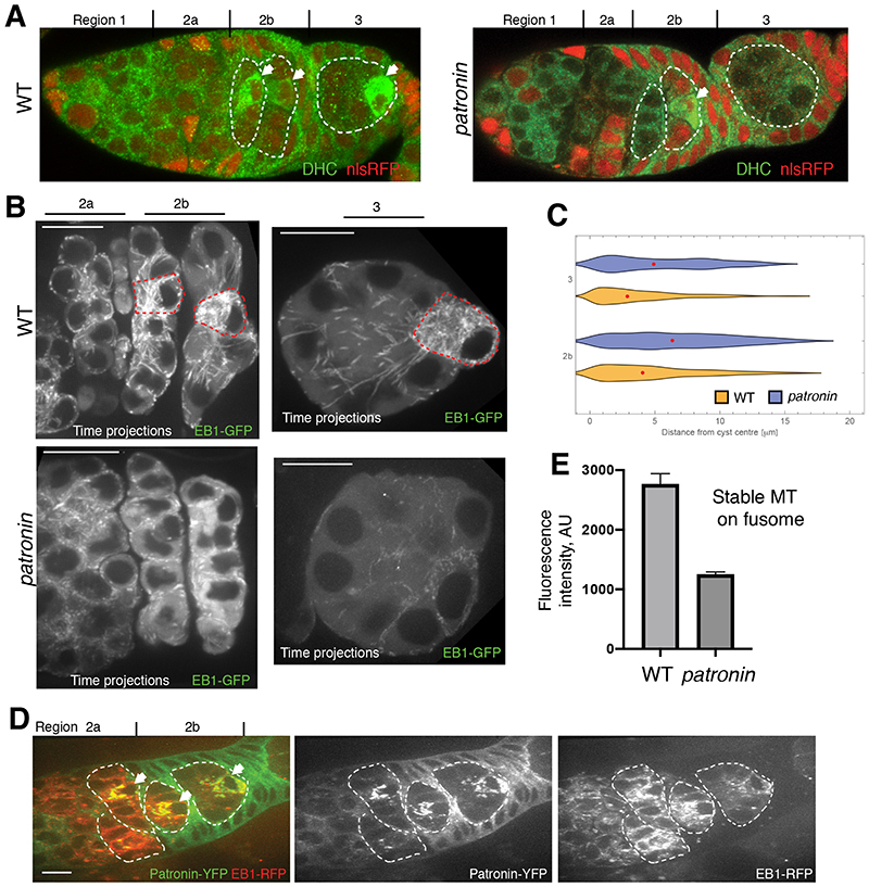
Fig. 3. Patronin is required for MT organisation in the cyst.
(A) Distribution of Dynein Heavy Chain (DHC) in wild type (WT) and patronin mutant cysts. (B-D) Patronin is required for MTOC formation in the presumptive oocyte. (B) EB-1 comet tracks in wild type (WT; top) and patronin mutant (bottom) cysts. The images are projections of several time points from Movies S1 (WT; region 2), S2 (WT; region 3), S3 (patronin; region 2) and S4 (patronin; region 3). The red dashed line marks cells with MTOCs. (C) Quantification of EB-1 comet distribution in wild type (WT) and patronin mutant cysts in region 3 and 2b of germarium. Red dots indicate median values. (D) Live germarium showing co-localization of Patronin-YFP foci with the microtubules plus end marker EB1-GFP in the presumptive oocyte. (E) Quantification of the mean fluorescence intensities of fusome associated acetylated microtubules in patronin mutant and WT cysts. Errors bars indicate the SEM.

