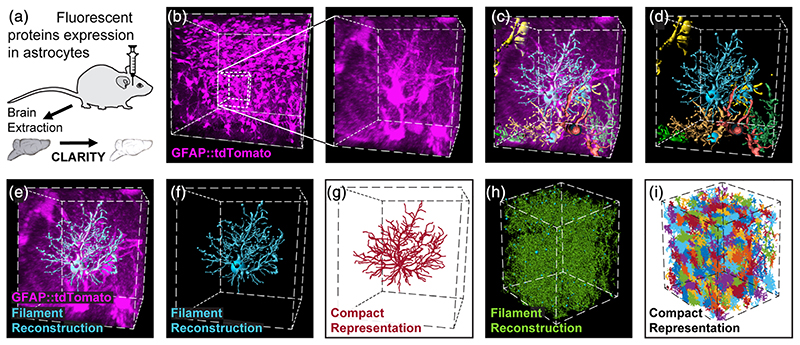
Figure 1. Automatic detection and reconstruction of astrocytic domains in large 3D volumes.
(a) Wild type or transgenic mice were injected with viral vectors to induce the expression of fluorescent proteins in astrocytes. Following >4 weeks, their brains were extracted, cut into thick hippocampal slices (4–5 mm) and were then made transparent by CLARITY after which they were imaged. (b) Expression of tdTomato (purple) in hippocampal astrocytes is presented for a 450 × 450 × 450 μm cube (left), and a zoomed-in 80 × 80 × 80 μm cube excerpt from it (right). (c, d) Seven representative astrocyte processes reconstructions from this cube are shown (and see Movie S1). Each astrocyte reconstruction, like the one shown in (e, f) was compactly represented (crimson) for further analysis (g). The process of processes reconstruction (green) (h) and compact representation (multicolor) (i) was performed on the entire imaging 500 × 400 × 400 μm cube

