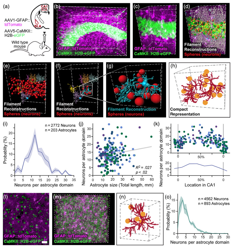
Figure 3. Automatic detection of excitatory neurons content and its distribution in astrocytic domains.
(a) The hippocampus and amygdala of mice were injected with viral vectors to induce the expression of the red fluorophore tdTomato in astrocytes, and the green fluorophore H2B-eGFP in pyramidal neurons nuclei in the hippocampus or amygdala, and thick slices (4-5 mm) were then rendered transparent by CLARITY. (b) Expression of tdTomato (purple) in hippocampal astrocytes and H2B-eGFP in pyramidal neurons (green) is presented fora 520 × 370 × 520 μm cube, (c) and a zoomed-in 250 × 120 × 150 μm cube. (d) All astrocyte processes reconstructions from this cube are shown (white, and see Movie S2), together with all neuronal somata (red). (e) Six representative filaments (blue, yellow, orange, pink, purple, and gray) of astrocytes crossing the pyramidal layer are shown with all neuronal somata (red). (f) The same six filaments are shown only with the neuronal somata (red) associated with the blue astrocyte. (g) A zoomed-in 80 × 70 × 100 μm cube showing the same reconstructed astrocyte and its associated neurons. (h) A compact representation of the same astrocyte (crimson) and its associated neurons (yellow). (i) The binned distribution (bin size = 2) of pyramidal neurons content of CA1 astrocytes (average = 13.7; n = 2772 neurons in 203 astrocytic domains in 4 cubes). Average presented in bold blue, with SEM shading. (j) A significant, albeit weak, positive correlation is found between the total processes length of the astrocyte and the number of pyramidal neurons in its domain (203 astrocytic domains, each represented by a dot, from n = 4 cubes, each displayed in a different color). (k) The number of pyramidal neurons per astrocytic domain increases toward the middle of the pyramidal layer. Average number (bottom) presented in bold blue, with SEM shading. (l) Expression of tdTomato (purple) in BLA astrocytes and H2B-eGFP in pyramidal neurons (green) is presented for a single slice (m) and a 290 × 250 × 130 μm cube. (see Movie S3). (n) A compact representation of an astrocyte (crimson) and its associated neurons (yellow). Cube volume: 90 × 80 × 60 μm. (o) The distribution of pyramidal neurons content of astrocytes in the BLA (average = 5.2; n = 4562 neurons in 893 astrocytic domains in three cubes). Average presented in bold light green, with SEM shading. Scale bar: 50 μm

