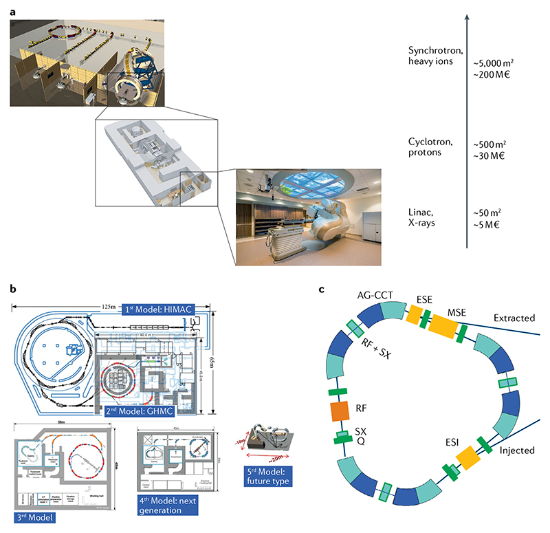
Figure 4. Accelerator technologies in heavy ion therapy.
a | The impact of advancing technology on footprint and costs of radiotherapy facilities. Size and prices can have large variation, but the image gives an indication of the increase in footprint and price. b | Plans at the National Institutes for Quantum and Radiological Science and Technology (NIRS-QST, Chiba, Japan) for reducing the footprint of heavy ion centres. The image shows the large research Heavy-Ion Medical Accelerator (HIMAC) and the subsequent concepts already implemented (such as the Gunma facility) or under development. GHMC, Gunma University Heavy Ion Medical Center. Image courtesy of K. Noda, NIRS-QST. c | An innovative concept of a superconducting synchrotron for heavy ion therapy developed at CERN. The triangular accelerator with a 3.5-T superconducting magnet is capable of accelerating C ions up to 430 MeV/n with 1010 ions per pulse in a footprint approximately one-quarter of the present-day resistive magnet medical synchrotrons in part b. AG-CCT, alternating gradient canted cosine theta; ESE, electrostatic extraction septum; ESI, electrostatic injection septum; MSE, magnetic extraction septum; Q, quadrupole magnet; RF, radiofrequency cavity; SX, sextupole magnet. Image and information courtesy of Elena Benedetto, CERN & TERA Foundation. Part a with permission from © GSI Helmholtzzentrum für Schwerionenforschung GmbH. Part b, image courtesy of Dr Koichi Noda. Part c, image courtesy of Elena Benedetto, TERA Foundation and CERN.

