. 2021 Oct 8;81(23):6004–6017. doi: 10.1158/0008-5472.CAN-21-1499

Figure 2.

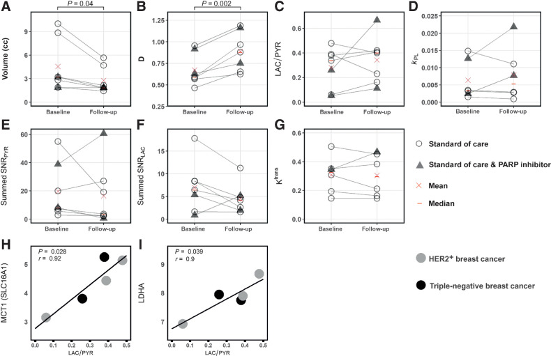
Figure 2. Parameters obtained from hyperpolarized 13C-MRI and 1H-MRI at baseline and in early follow-up scans. Differences between baseline and follow-up were significant for tumor volume (A) and diffusivity (B) but not for the other parameters (C–G); neither change in volume or diffusivity could distinguish pCR from non-pCR. Correlation of SLC16A1 (MCT1) and LDHA mRNA expression with LAC/PYR was significant (H and I). Only images acquired with identical 13C-MRI acquisition parameters (spectral–spatial excitation) were included in these correlations.

Parameters obtained from hyperpolarized 13C-MRI and 1H-MRI at baseline and in early follow-up scans. Differences between baseline and follow-up were significant for tumor volume (A) and diffusivity (B) but not for the other parameters (C–G); neither change in volume or diffusivity could distinguish pCR from non-pCR. Correlation of SLC16A1 (MCT1) and LDHA mRNA expression with LAC/PYR was significant (H and I). Only images acquired with identical 13C-MRI acquisition parameters (spectral–spatial excitation) were included in these correlations.