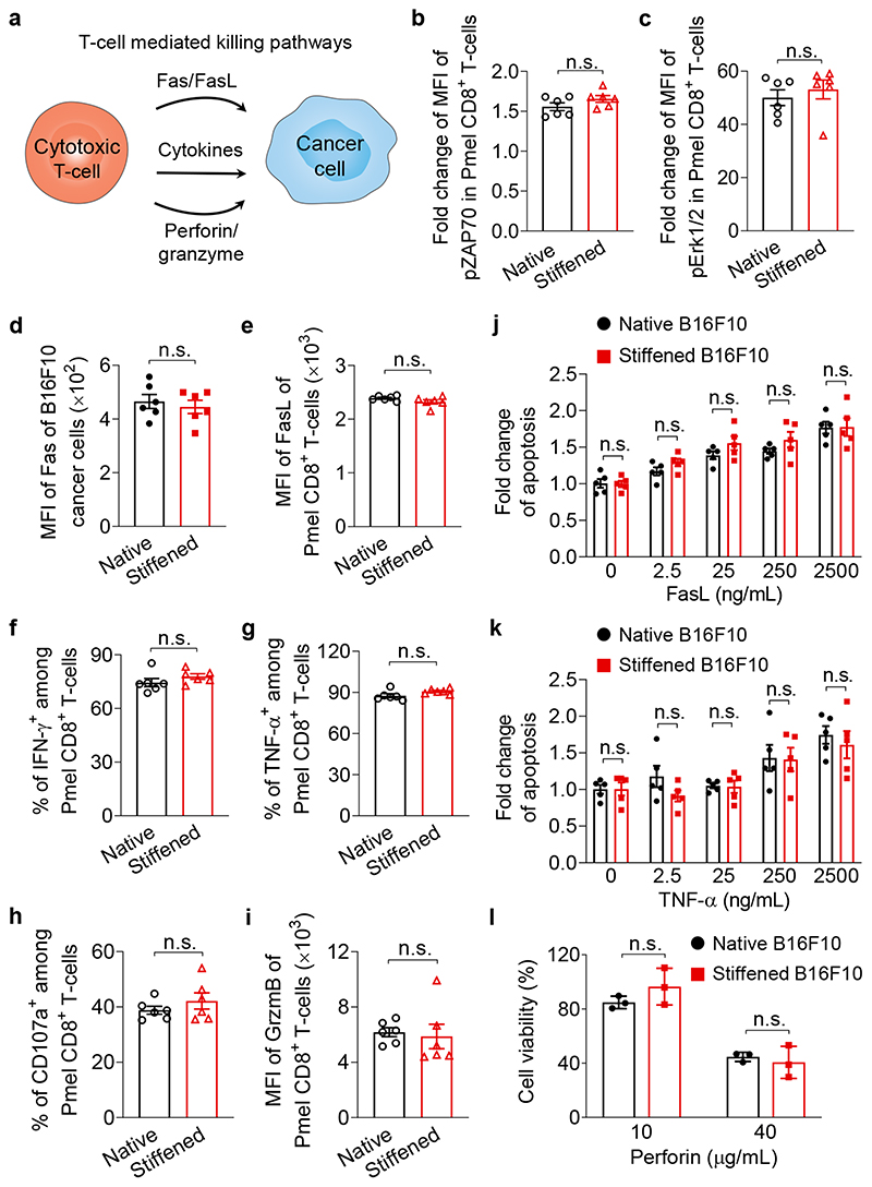
Fig. 5. Cancer-cell stiffening has negligible influence on biochemical cancer-cell killing pathways mediated by T-cells.
a, Schematic illustration of T-cell mediated killing pathways. b, c, Fold change of MFI of phosphorylated ZAP70 (pZAP70, b) and Erk1/2 (pErk1/2, c) in activated Pmel CD8+ T-cells stimulated by native or MeβCD-treated (stiffened) B16F10 cancer cells at 37 °C for 5 min (n = 6). d, Expression levels of Fas of native and stiffened B16F10 cancer cells (n = 6). e-i, Activated Pmel CD8+ T-cells were co-cultured with native or stiffened B16F10 cancer cells (E:T ratio = 10:1) at 37 °C for 5 h. Shown are expression levels of Fas ligand (FasL) (e) and granzyme B (GrzmB) (i), and frequencies of IFN-γ+ (f), TNF-α+ (g), and CD107a+ (h) of Pmel CD8+ T-cells (n = 6). j, k, Fold change of frequencies of apoptotic native and stiffened B16F10 cancer cells after incubation with FasL (j) or TNF-α (k) at indicated concentrations at 37 °C for 5 h (n = 5). l, Viability of native and stiffened B16F10 cancer cells after incubation with perforin of indicated concentrations at 37 °C for 20 min (n = 3). P values were determined by unpaired Student’s t test. Error bars represent SEM. MFI, mean fluorescence intensity; n.s., not significant. All data are one representative of at least three independent experiments with biological replicates.

