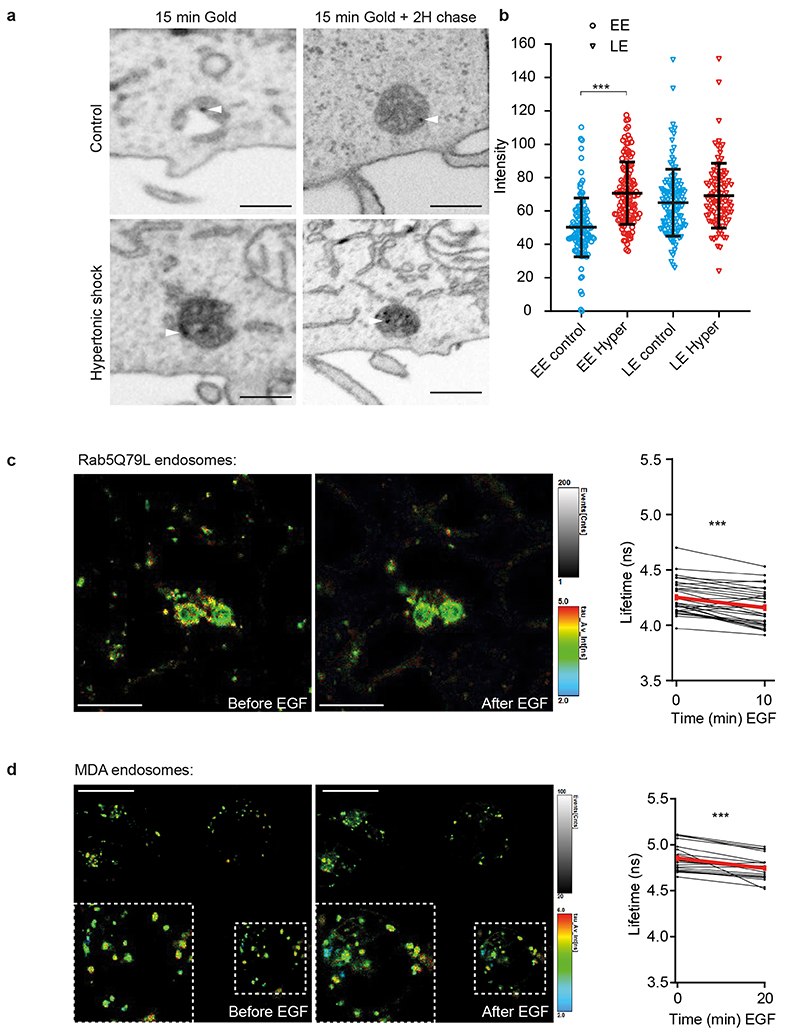
Extended Data Fig. 8. EGF triggers endosomal membrane tension decrease in vivo (related to Fig 6).
a-b) As in Fig 4, the panel (a) shows FIB-SEM micrographs of cells loaded with BSA-gold. The electron density of early endosomes (EE) or late endosomes (LE) labeled with BSA-gold was measured before (blue) and after (red) a 10min hypertonic shock (b). Mean±SD (N are respectively EE control:121; EE Hyper: 124; LE control:120; LE Hyper:98 from 2 independent replicates, Kruskal-Wallis test: P<1.10-15). c) Lyso Flipper lifetime measurements on RAB5Q79L endosomes before and 10min after 200 ng/ml EGF treatment. Thin lines: independent images from 4 experiments, thick red line: mean±SEM, two-tailed paired t-test P= 0.0000025902. d) Lyso Flipper lifetime measurements in MDA cells before and 20min after 200 ng/ml EGF treatment. Thin lines: independent images from 5 experiments, thick red line: mean±SEM, two-tailed paired t-test P=0.0000496474.

