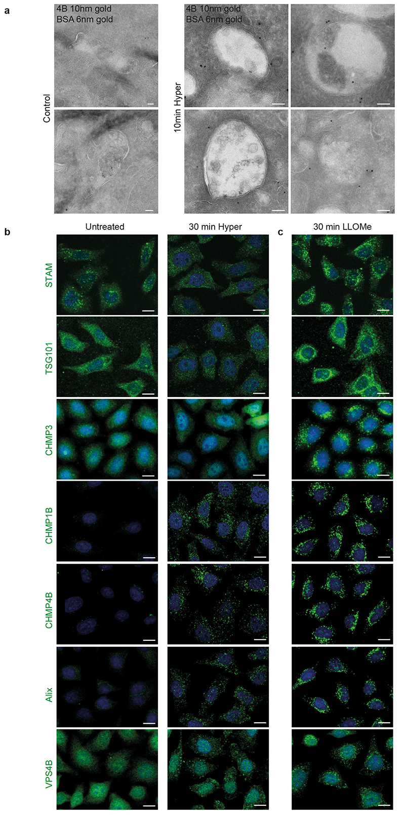
Extended Data Fig. 2. Hypertonic shock or LLOMe treatment leads to ESCRT proteins relocalization (related to Fig 1–2 & 4).
a) Hela-CHMP4B-GFP cells were incubated for 15min with 6nm BSA-Gold and subjected to a hypertonic shock for 10min. and cells were processed for immune-electron microscopy, as in Fig 1k. The gallery shows representatives micrographs of endosomes after immuno-gold labelling of cryo-sections with anti-CHMP4B antibodies followed by 10 nm Gold-Protein A. Please note that the size of endosomes treated or not under hypertonic conditions cannot be compared in these micrographs. Indeed, we were unable to obtain nice thin cryo-sections after fixation in a paraformaldehyde-glutaraldehyde mixture containing sucrose (in order to keep osmolarity constant). Thus, we had to use normal fixative to obtain appropriate sections, but some endosome swelling occurred. Images from a single immuno-gold labelling experiment. b-c) Representatives automated confocal images of Hela MZ cells after immunostaining of the indicated ESCRT subunits under the following conditions: (b) untreated and 30min hypertonic shock, (c) 30min treatment with LLOMe. Quantifications in Fig 1l and Fig 2n were obtained from similar images. Images representative of 3 experiments. Scale bars: 10μm

