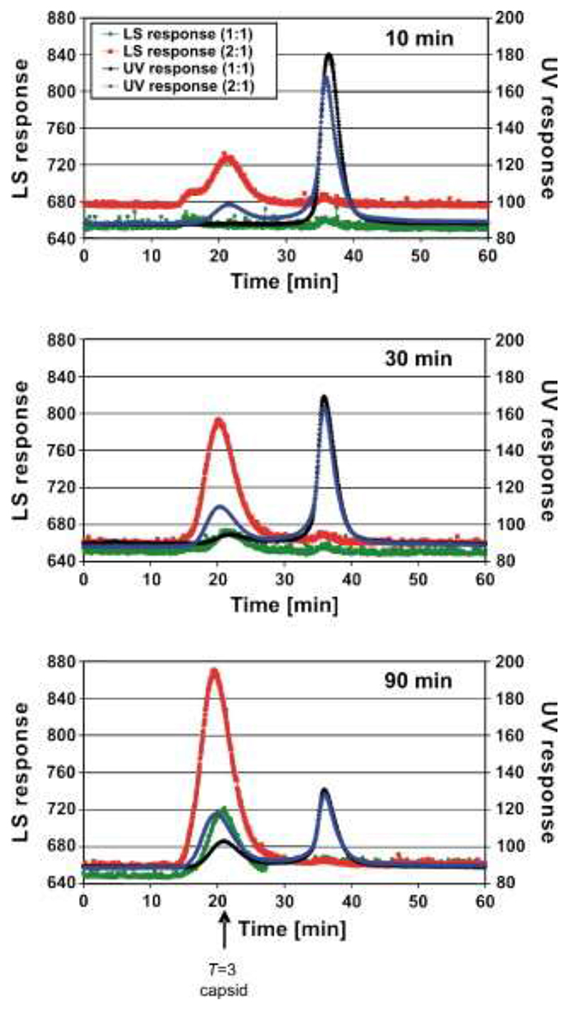
Figure 3.
Gel filtration-light-scattering assays of capsid reassembly. MS2 CP2 and TR were mixed in 40 mM ammonium acetate, pH 5.2−5.7, to form a 1:1 reaction, incubated at 4 °C for 10, 30, or 90 min, and then loaded onto a Sepharose 6 gel filtration column equilibrated in 50 mM Tris−acetate, pH 7.4, and eluted at 0.36 ml/min. The outflow from the column was analyzed simultaneously via UV absorbance (black trace) and light-scattering (green trace). Similar reactions were then set up, preincubated at a molar ratio of 1:1 for 10 min, and then an additional aliquot of protein was added to create the 2:1 reaction. The traces are shown as UV absorbance (blue trace) and light-scattering (red trace). The position at which authentic T = 3 capsids elute is marked with an arrow; the peak at ∼35 min corresponds to the starting materials and smaller complexes.

