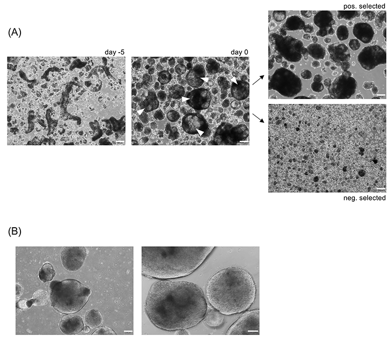
Figure 3. Morphology of primed aggregates.
(A) Colony fragments of hCD34iPSC16 (left picture, day -5) were cultured in 6-well suspension culture plate on an orbital shaker in priming medium for 5 days. White error heads indicate primed aggregates with optimal morphology which is defined by a size of > 300-500 μm diameter, the presence of some cystic structures within the aggregates and a translucent appearance (middle picture, day 0). The largest primed aggregates were selected by defined sedimentation. Upper image shows larger, positively selected primed aggregates; lower image shows smaller, negatively selected aggregates. Scale bar: 200 μm, magnification 40x. (B) Primed aggregates with sub-optimal morphology appearing round and dense. Scale bar 200 μm, 40x magnification and 100 μm, 100x magnification.

