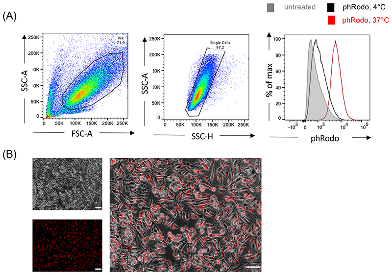
Figure 8. Phagocytosis of bioparticles by iPSC-derived macrophages (related to BOX 5).
To evaluate the functionality of iPSC-Mac, cells were incubated with pHrodo™ Red E. coli BioParticles™ for 4 hours at 4 or 37 °C. (A) Flow cytometric analysis. Pre-gating on viable cells by FSC/SSC properties was followed by a single-cell gating step (SSC-A vs. SSC-H). Untreated iPSC-Mac (grey filled, 143.913 cells), iPSC-Mac incubated with bioparticles at 4 °C (black line, 100.785 cells) and iPSC-Mac incubated with bioparticles at 37 °C (70.466 cells) were analyzed. Flow cytometry was performed with a Cytoflex S (Beckman Coulter) and data was analyzed using FlowJo (BD Bioscience). Underlying flow cytometry data sets can be accessed under http://flowrepository.org/, Repository IDs: FR-FCM-Z4WC). (B) Fluorescence microscopy of iPSC-Mac incubated with pHrodo™ Red E. coli BioParticles™ at 37 °C (scale bar: 100 μm, magnification 200x).

