. 2021 Jul 19;11(12):3158–3177. doi: 10.1158/2159-8290.CD-21-0209

Figure 1.

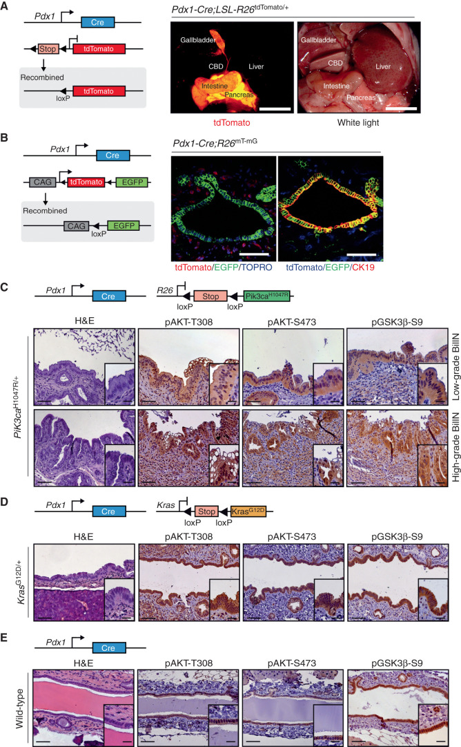
Figure 1. Constitutive activation of the PI3K signaling pathway induces premalignant biliary intraepithelial neoplasia (BilIN). A, Left: genetic strategy and recombination scheme to analyze the patterns of Pdx1-Cre transgene expression using a conditional tdTomato reporter allele (LSL-R26tdTomato/+). Right: macroscopic fluorescence and white-light images of the Pdx1-Cre;LSL-R26tdTomato/+ reporter mouse. Visualization of tdTomato reveals reporter gene expression (red) in the pancreas, duodenum, gallbladder, and common bile duct (CBD). Scale bars, 1 cm. B, Left: genetic strategy and recombination scheme to analyze the patterns of Pdx1-Cre transgene expression using a switchable floxed double-color fluorescent tdTomato-EGFP Cre reporter line (R26mT-mG). Right panel, left image: representative confocal microscopic image of tdTomato (red color, non–Cre-recombined cells) and Cre-induced EGFP (green color) expression in the common bile duct. Note: Cre-mediated EGFP expression in the biliary epithelium and peribiliary glands, but not stromal cells. Nuclei were counterstained with TOPRO-3 (blue). Right image: representative immunofluorescence staining of CK19 (red color) of Pdx1-Cre;R26mT-mG/+ animal. Note: Blue color shows expression of tdTomato in unrecombined cells. Green color labels Cre-recombined cells that express EGFP. Colocalization of CK19 immunofluorescence staining (red) and EGFP expression (green) results in yellow color. Scale bars, 50 μm. C–E, Genetic strategy used to express oncogenic Pik3caH1047R (C), KrasG12D (D), or only Cre as control (E) in the common bile duct (top). Hematoxylin and eosin (H&E) staining and IHC analysis of PI3K/AKT pathway activation in the biliary epithelium of the common bile duct and different grades of dysplasia in BilIN (bottom). Scale bars, 50 μm for micrographs and 20 μm for insets.

Constitutive activation of the PI3K signaling pathway induces premalignant biliary intraepithelial neoplasia (BilIN). A, Left: genetic strategy and recombination scheme to analyze the patterns of Pdx1-Cre transgene expression using a conditional tdTomato reporter allele (LSL-R26tdTomato/+). Right: macroscopic fluorescence and white-light images of the Pdx1-Cre;LSL-R26tdTomato/+ reporter mouse. Visualization of tdTomato reveals reporter gene expression (red) in the pancreas, duodenum, gallbladder, and common bile duct (CBD). Scale bars, 1 cm. B, Left: genetic strategy and recombination scheme to analyze the patterns of Pdx1-Cre transgene expression using a switchable floxed double-color fluorescent tdTomato-EGFP Cre reporter line (R26mT-mG). Right panel, left image: representative confocal microscopic image of tdTomato (red color, non–Cre-recombined cells) and Cre-induced EGFP (green color) expression in the common bile duct. Note: Cre-mediated EGFP expression in the biliary epithelium and peribiliary glands, but not stromal cells. Nuclei were counterstained with TOPRO-3 (blue). Right image: representative immunofluorescence staining of CK19 (red color) of Pdx1-Cre;R26mT-mG/+ animal. Note: Blue color shows expression of tdTomato in unrecombined cells. Green color labels Cre-recombined cells that express EGFP. Colocalization of CK19 immunofluorescence staining (red) and EGFP expression (green) results in yellow color. Scale bars, 50 μm. C–E, Genetic strategy used to express oncogenic Pik3caH1047R (C), KrasG12D (D), or only Cre as control (E) in the common bile duct (top). Hematoxylin and eosin (H&E) staining and IHC analysis of PI3K/AKT pathway activation in the biliary epithelium of the common bile duct and different grades of dysplasia in BilIN (bottom). Scale bars, 50 μm for micrographs and 20 μm for insets.