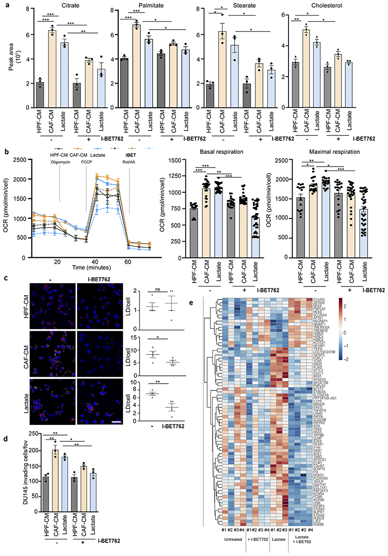
Figure 4. Lactate-induced metabolism and invasiveness are sensitive to BET inhibition in PCa cells.
a) Citrate, palmitate, stearate and cholesterol levels were detected in DU145 cells treated with HPF- or CAF-CM and 15mM lactate for 48h, ± I-BET762 (100nM). One-way ANOVA; Tukey’s corrected; *p < 0.05; **p < 0.01; ***p < 0.001 b) OCR from MitoStressTest analysis performed on DU145 cells treated as indicated ± I-BET762. Data are represented as mean ± SEM of three independent experiments (≥ 4 technical replicates). c) BODIPY 493/503 staining of DU145 cells treated as indicated ± I-BET762. Quantification of BODIPY spots per cell was reported. Scale bar: 10 μm d) Invasion assay of DU145 cells treated as indicated ± I-BET762. e) Heatmap showing the expression of differentially expressed genes between DU145 cells exposed to 15mM lactate for 48h and treated w/wo I-BET762.

