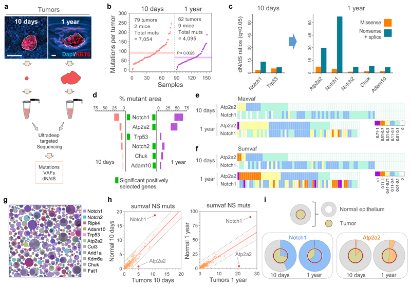
Figure 2. Sequencing of tumors and adjacent normal tissue.
(a) Esophageal tumors were collected 10-days or 1-year post-Diethylnitrosamine (DEN) treatment and ultra-deep targeted sequencing with a 192 gene bait-set performed. Scale-bars: 50μm. (b) Mutations per tumor, including essential splice, frameshift, missense, nonsense and silent mutations. Dotted lines and colored shadows indicate mean±s.e.m (two-tailed Mann-Whitney test). (c) dN/dS ratios for missense and truncating (nonsense + essential splice site) mutations of positively selected genes (dN/dS>1) in the 10 days and 1 year post-DEN tumors (only significant genes for each time point are shown, q<0.05 calculated with R package dNdScv26). (d) Estimated percentage of tumor area carrying non-synonymous mutations in the positively selected genes (identified with green rectangles for each time point). Range indicates upper and lower bound estimates. (e, f) Heatmaps indicating the maximum (e) and summed VAF (f) for non-synonymous mutations in Atp2a2 and Notch1 in 10-day and 1-year post-DEN tumors. (g) Schematic representation of the mutant clones in normal esophageal epithelium collected 10 days post-DEN treatment. Only mutations of the positively selected genes (dN/dS>1) in normal or tumor samples at 10 days and 1 year13 are shown. The density and size of the clones are inferred from the sequencing data, and the nesting of clones and subclones is inferred from the data when possible and randomly allocated otherwise. (h) Correlations between the summed VAF of nonsynonymous mutations (NS muts) in the 192 genes sequenced from normal epithelium and tumors at 10-days (left) and 1-year (right) post-DEN treatment. Centre lines show two-tailed Pearson correlations (R2=0.8329; P=1.6x10-76) and R2=0.7081; P=3.2x10-53, respectively). Red dots indicate genes outside the 99% predicted band (colored area within grey dotted lines). (i) Percentage of area covered by Notch1 (blue, left graphs) and Atp2a2 (orange, right graphs) mutations in the normal epithelium (outer circles) versus the tumors (inner circles) collected at 10-days or 1-year post-DEN treatment.

