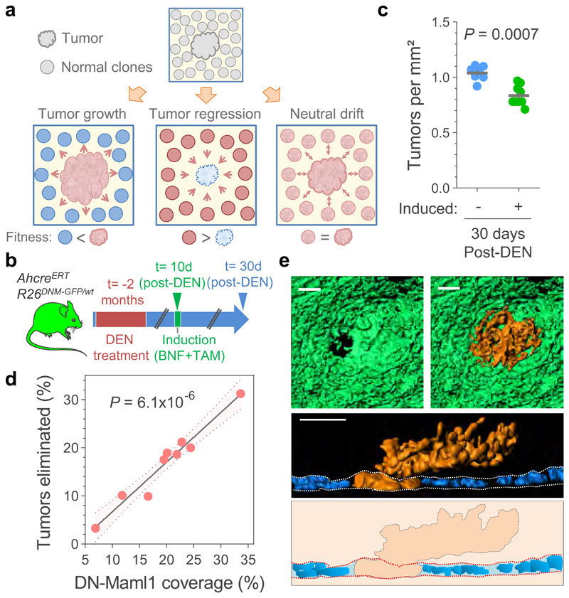
Figure 3. Elimination of early tumors by expansion of mutant clones in the adjacent normal epithelium.
(a) Hypothesis: Tumor survival depends on their competitive fitness relative to that of the surrounding normal tissue, predicting 3 possible scenarios: 1) tumor cells are fitter than clones in the normal adjacent epithelium (left), tilting the balance towards tumor growth; 2) Tumor cells are less fit than the adjacent normal epithelium (middle), resulting in tumor regression and elimination, and 3) cells in tumor and adjacent normal epithelium are equally fit (right), resulting in neutral drift. (b) Protocol: AhcreERT/R26DNM–GFP/wt mice received DEN for two months. 10 days after DEN withdrawal DN-Maml1 clones were induced and tissues harvested twenty days later. Control mice received DEN but were not induced. (c) Number of tumors per mm2 of esophageal epithelium in induced and non-induced AhcreERT/R26DNM–GFP/wt mice (n= 9 and 7 mice, respectively). Lines indicate mean±s.e.m (two-tailed Mann-Whitney test). (d) Correlation between the area covered by DN-Mam1 clones and tumors being eliminated (see Methods). n=9 mice. Line shows the two-tailed Pearson correlation (R2=0.9541) with 95% confidence interval (colored area). (e) Rendered confocal images showing top views (top images), lateral view (middle image) and schematic (bottom image) of a tumor (orange) surrounded by a DN-Maml1 clone (green). Dapi is shown in blue. Scale-bars: 20μm.

