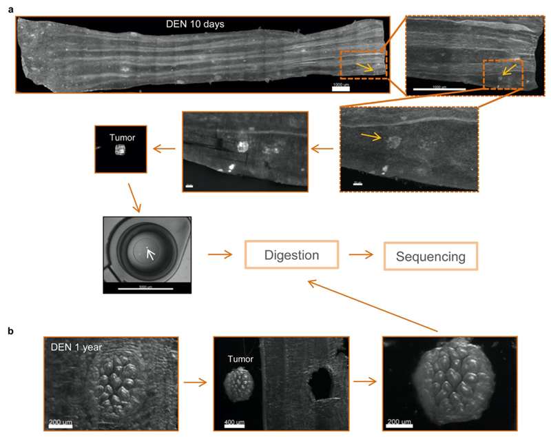
Extended Data Figure 3. Collection of 10-day and 1-year esophageal tumors.
(a-b) Mouse esophagus was collected 10-days (a) or 1-year (b) post-DEN treatment. The esophagus was cut open longitudinally and the epithelium separated from the underlying muscle and stroma. The epithelium was flattened, fixed, stained with KRT6 (grey), mounted and 3D-imaged on a confocal microscope. Tumors were identified from the processed images and manually dissected under a fluorescent microscope and sequenced (see Methods).

