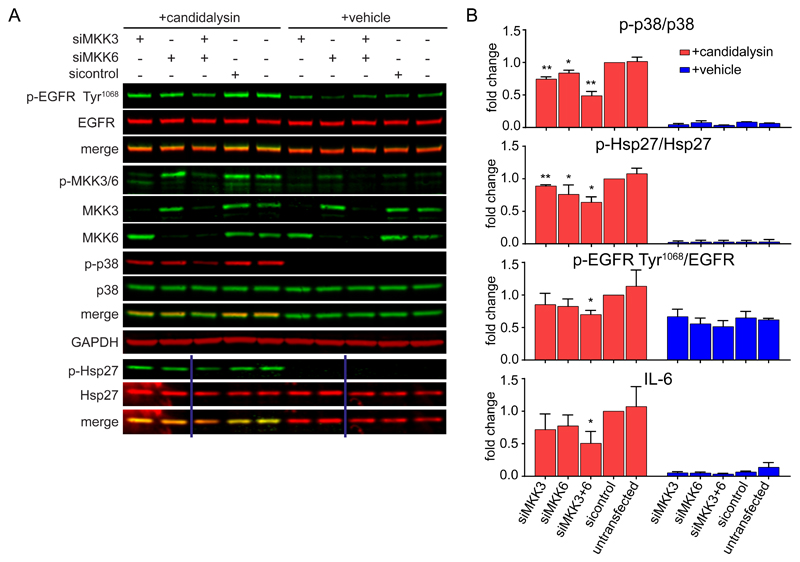
Fig. 4. MKK3 and MKK6 trigger candidalysin-induced p38 signaling.
A, Representative immunoblot showing phosphorylation (p-) of EGFR on Tyr1068, MKK3/6, p38, and Hsp27 and total MKK3 and MKK6 protein in TR146 OEC cells transfected for 72 h with siRNAs for MKK3 and/or MKK6 prior to candidalysin stimulation for 30 min. Immunoblots are representative of three biological replicates. GAPDH is a loading control. Vertical lines represent intervening gel lanes that are not shown. B, Graphical quantification of immunoblots as in A and IL-6 release from TR146 cells transfected for 72 h with siRNA for MKK3 and/or MKK6 prior to candidalysin stimulation for 24 h. Graphs show means of three biological replicates + SD and are expressed as fold change relative to siRNA control + candidalysin. Statistical significance was quantified by one sample t test compared to a hypothetical value = 1. *P < 0.05, **P < 0.01.

