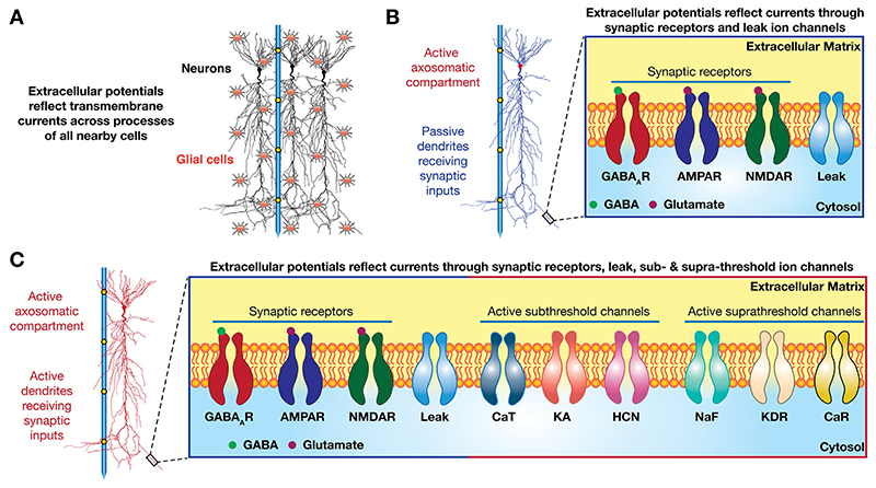
Fig. 1. Sources of LFP.
(A) A schematic of LFP recording from a population of principal neurons and astrocytes using a single electrode with multiple contact points that serve as recording sites. (B) A single recording electrode with multiple sites and a single pyramidal neuron passive dendrites and voltage-gated ion channels only in the axo-somatic compartments. A small dendritic segment is expanded to highlight various synaptic receptors and passive leak channels that contribute to the transmembrane currents. These currents are in addition to the capacitive current consequent to the two ion-conducting media (the cytosol and the cerebrospinal fluid) separated by a dielectric lipid bilayer. (C) Same as B but with active dendrites. An expanded view of a small dendritic segment highlights the diversity of sub- and supra-threshold ion channels/conductances that also contribute to the transmembrane currents. The morphological reconstructions are modified from neuron n123 in the open-source neuronal morphology database Neuromorpho.org (Ascoli et al., 2007).

