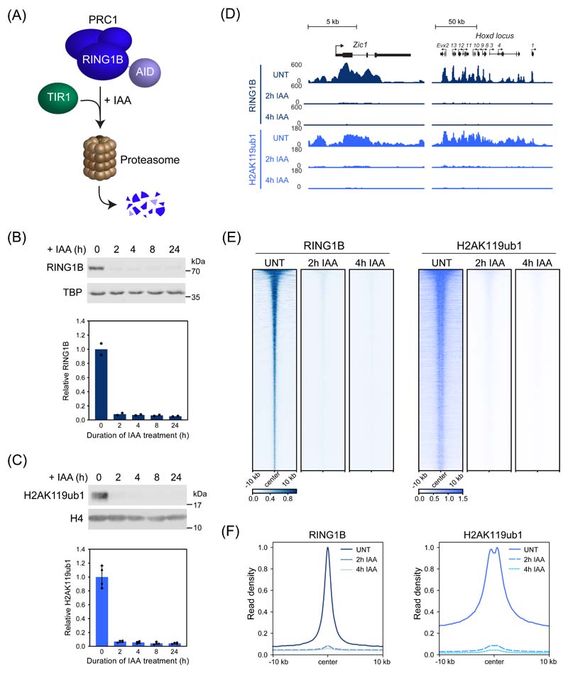
Figure 1. Acute depletion of PRC1 reveals a rapid turnover of H2AK119ub1.
(A) A schematic illustrating the PRC1deg system. Addition of auxin (IAA) induces proteasomal degradation of AID-RING1B.
(B) Western blot analysis (upper panel) and quantification (lower panel) of RING1B in PRC1deg cells treated with IAA for the indicated times. Shown are values and mean from n=2 independent experiments.
(C) Western blot analysis (upper panel) and quantification (lower panel) of H2AK119ub1 in PRC1deg cells treated with IAA for the indicated times. Shown are values and mean ± SD from n=4 independent experiments.
(D) Genomic snapshots of typical Polycomb target genes, showing cChIP-seq signal for RING1B and H2AK119ub1 in PRC1deg cells treated with IAA for the indicated times.
(E) Heatmap analysis of RING1B (left) and H2AK119ub1 (right) cChIP-seq at RING1B-bound sites in PRC1deg cells treated with IAA for the indicated times. Heatmaps were sorted by RING1B signal in untreated cells.
(F) Metaplot analysis of data shown in (E). Maximal read density in untreated cells was set to 1.

