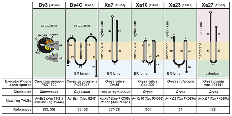
Figure 5. Structural and functional of executor R genes and encoded executor proteins.
Designation of executors is given on top with the size of the executor protein in square brackets. Depictions display the predicted topology and subcellular localization of executor proteins with terminal white and black circles indicating N- and C-termini, respectively. Transmembrane stretches were predicted with TMHMM (http://www.cbs.dtu.dk/services/TMHMM-2.0/) and are indicated with grey-coloured lines. Digits indicate the number of amino acids domains that a given domain is composed of.

