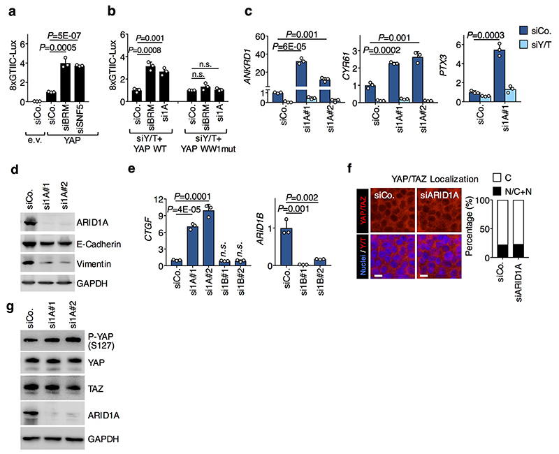
Extended Data Figure. 2. Effect of ARID1A depletion on YAP/TAZ levels, localization and activity.
a, The panel represents the results of luciferase assays with the 8xGTIIC-Lux reporter in HEK293 cells transfected with empty (e.v.) or YAP-expressing vectors and the indicated siRNAs. Data are normalized to siCo/empty vector-transfected cells and are presented as mean + s.d. of n=3 biologically independent samples.
b, The panel represents the results of luciferase assays with the 8xGTIIC-Lux reporter in HEK293 cells reconstituted with either YAP wt or YAP WW1mut and transfected with the indicated siRNAs. Data are normalized to siCo-transfected cells and are presented as mean + s.d. of n=3 biologically independent samples.
c, Panels are qRT-PCR analyses for the YAP/TAZ targets ANKRD1, CYR61 and PTX3 (mean + s.d. of n=3 biologically independent samples) in MCF10A cells transfected as indicated.
d, Western blot analysis for ARID1A, E-Cadherin and Vimentin from lysates of MCF10A cells transfected with the indicated siRNAs.
e, Panels are qRT-PCR analyses for CTGF (left) and ARID1B (right) expression (mean + s.d. of n=3 biologically independent samples) in MCF10A cells transfected as indicated.
f, Representative confocal images (left) and quantifications (right; >100 cells per condition) of YAP/TAZ localization in MCF10A cells transfected with the indicated siRNAs.
g, Western blot analysis for YAP, TAZ and YAP phosphorylation on the key Hippo/LATS target site (P-YAP S127) from lysates of MCF10AT cells transfected with the indicated siRNAs.
P values were determined by unpaired two-sided t-test.
All panels display representative experiments, repeated independently two (d, e, g) or three (a-c, f) times with similar results.

