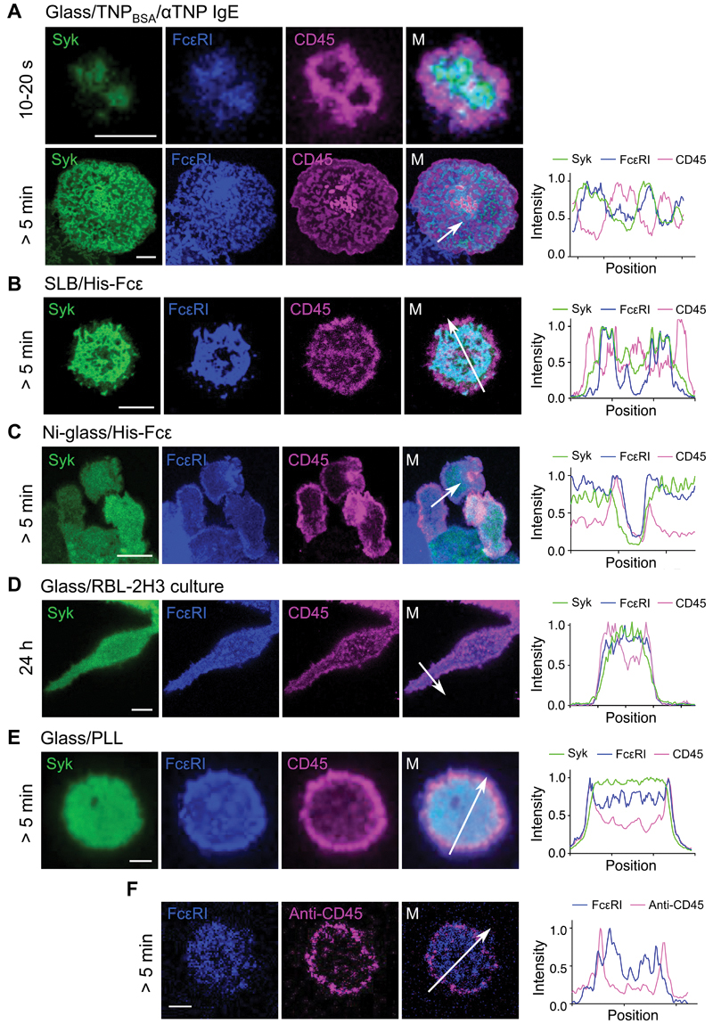
Fig. 3. CD45 and FcεRI segregate following engagement of surface-associated IgE.
(A to E) Left: Confocal fluorescence images of Syk, FcεRI, and CD45 at the basal surfaces of RBL-2H3 cells that had made contact with (A) TNPBSA-IgE-coated glass, (B) a His-Fcε-functionalized SLB, (C) Ni-glass, (D) uncoated glass, or (E) PLL-coated glass. Timings indicate the period of contact with the surface. Right: Intensity line profiles correspond to the arrows in the merged views. (F) Exclusion of CD45 relative to FcεRI was observed for primary human basophils that were loaded with anti-Der f 1 IgE and then interacted with Der f 1–coated glass. Images are representative of observations made in at least three independent experiments. Replicates with primary basophils were performed with blood from different donors. Scale bars, 5 µm.

