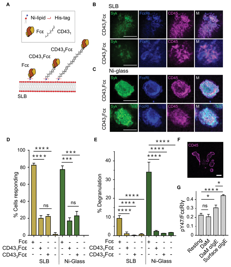
Fig. 5. CD45 exclusion is sensitive to the size of the surface-associated ligand.
(A) Schematic representation of CD43Fcε chimeras. (B and C) Confocal fluorescence images of Syk, FcεRI, and CD45 at the basal surface of an RBL-2H3 cell after making contact with an SLB (B) or Ni-glass (C) presenting extended CD431Fcε and CD433Fcε chimeras. (D) Percentages of RBL-2H3 cells producing Ca2+ fluxes within 10 min of making contact with an SLB or Ni-coated glass functionalized with extended CD431Fcε and CD433Fcε chimeras versus the same surfaces presenting His-Fcε. Data are means ± SD of four independent experiments. (E) Confocal fluorescence image of CD45 (that is, anti-CD45 Fab) at the basal surface of primary human basophils after making contact with anti-IgE–coated glass. (F) Normalized anti-pY47 FcεRIγ signals relative to anti-FcεRIγ signals obtained from Western blots of whole-cell lysates from primary human basophils after making contact anti-IgE conjugated directly or indirectly (through donkey anti-mouse; DaM) to glass. Data are means ± SD of three independent experiments. Resting denotes cells kept in suspension. (G) Percentage maximal degranulation of RBL-2H3 cells 30 min after making contact with an SLB or Ni-coated glass functionalized with extended CD431Fcε and CD433Fcε chimeras versus the same surfaces presenting His-Fcε. Data are means ± SD of four independent experiments. Images are representative of observations made in at least three independent experiments. Replicates with primary basophils were performed with blood from different donors. Scale bars, 5 µm. *P < 0.05, **P < 0.01, ***P < 0.001; ns, not significant.

