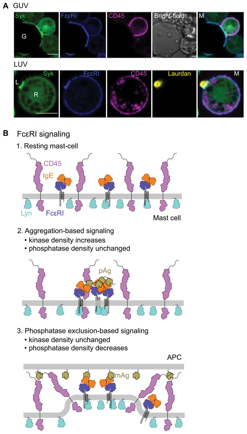
Fig. 7. FcεRI signaling can be triggered by unilamellar vesicles.
(A) Confocal fluorescence images of Syk, FcεRI, and CD45 at the equatorial plane of RBL-2H3 cells that interacted with GUVs (top) or laudan-stained LUVs (bottom) loaded with Fcε. G, GUV; L, LUV. Scale bars, are 5 µm. Images are representative of observations made in three independent experiments. (B) Schematic representation of the non-triggered state of the resting basophil surface (top) and after the triggering of FcεRI by polyvalent antigen (pAg; middle) or surface-associated monovalent antigen (mAg; bottom).

