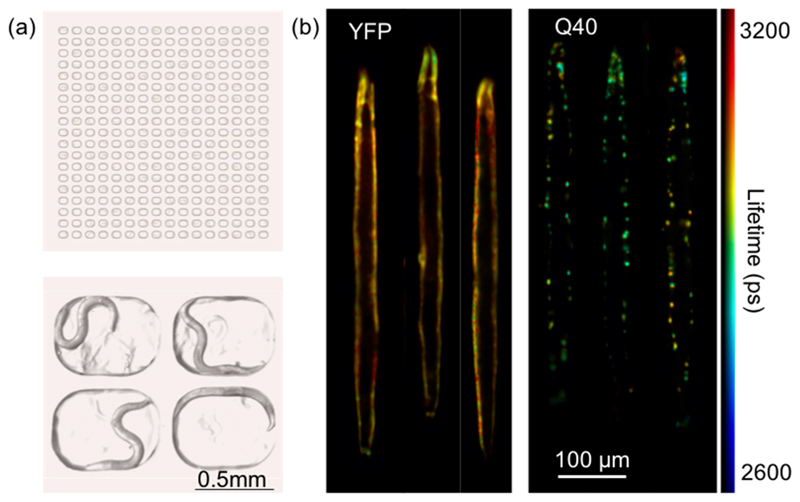
Figure 4. TG-FLIM applied to live C. elegans crawling in agarose microchambers.
(a) Schematic of the agarose-based microchamber device loaded with C. elegans (top) and an image of four chambers, each occupied by a single worm (bottom). (b) Registered and digitally stretched fluorescence lifetime maps of live crawling C. elegans. Shown here are example images of YFP and Q40 worms at day 3 of adulthood. For each worm, 10 sequential FLIM acquisitions were carried out (equaling a total acquisition time of ~5 s, therefore comparable to that used for anaesthetized worms ~4.2 s), and the fluorescence lifetime maps were averaged after digital stretching.

