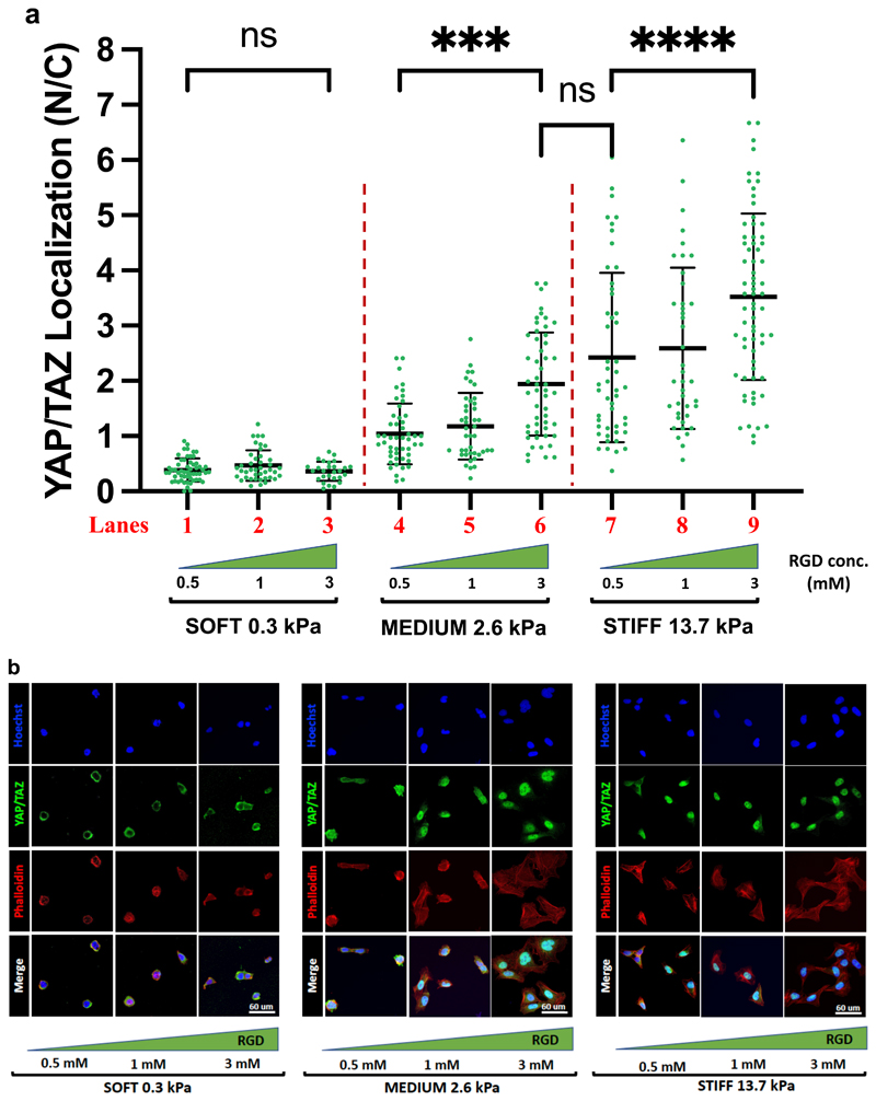
Figure 3.
Representative immunofluorescence images (a, c) and quantifications (b, d) of the nuclear/cytoplasmic (N/C) ratio of YAP/TAZ subcellular localization in MCF10A (a, b) or U2OS (c, d) cells, after seeding on PAA-OH substrates, coated with FN of five different stiffness. From the staining are visible: nuclei (in blue), F-actin (in red), YAP/TAZ (in green). F-actin was stained with fluorescently labeled phalloidin to serve as cell shape reference. Each dot in b) and d) corresponds to quantification of the N/C ratios of YAP/TAZ subcellular localization in individual cells, as obtained with Software-assisted Imaging processing of confocal images (see methods). Number of cells for each lanes are: b) lane 1: 116; lane 2: 93; lane 3: 146; lane 4: 210; lane 5: 120. d) lane 1: 36; lane 2: 27; lane 3: 31; lane 4: 48; lane 5: 48.

