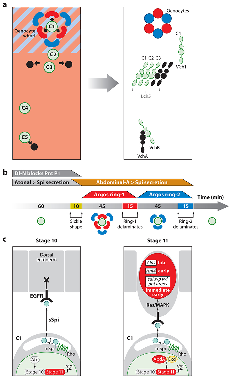
Figure 4. Oenocyte induction in Drosophila melanogaster.
(a) Fate map for oenocytes and chordotonal organs. Panels represent a single abdominal hemisegment at (left) early (stage 11) and (right) late (stage 16) embryogenesis. At stage 11, a whorl of approximately six sickle-shaped oenocyte precursors (red and blue) is induced by the most dorsal primary COP, C1 (green) from dorsoposterior ectoderm expressing Spalt major (light blue) and Engrailed (pink). More ventrally positioned primary COPs lying outside the Spalt domain (C3, C5, and possibly C2) induce secondary COPs (black) giving rise to chordotonal organs. By stage 16, all oenocytes and chordotonal organs have delaminated beneath the ectoderm and differentiated within their characteristic clusters. Adapted with permission from References 14, 15, and 41. (b) Embryonic timeline (minutes) showing two three-by-three pulses of oenocyte delamination during stage 11. Above the line, trapezoids indicate the duration of Atonal-dependent Spi secretion from C1 (gray), inhibition by Notch (dark gray), and AbdA-dependent secretion from C1 (orange) as well as the strong EGFR response in oenocyte precursors marked by Argos expression in ring-1 (red) and ring-2 (blue). (c) Model for oenocyte specification by AbdA. A primary COP (lower cell) signaling to the dorsal ectoderm via rbo-dependent Spi secretion is depicted. During stage 10, the EGFR response is blocked by Notch signaling and probably other factors. During stage 11, AbdA and Exd maintain rbo transcription, thus converting membrane-bound (mSpi) to secreted Spitz (sSpi), activating the EGFR and thus triggering immediate-early, early, and late gene expression in differentiating oenocytes. Abbreviations: AbdA, Abdominal-A; COP, chordotonal organ precursor; EGFR, epidermal growth factor receptor.

