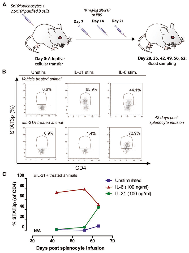
Figure 1. STAT3p is effectively blocked by αIL-21R.
A, Schematic representation of the treatment strategy to measure blockade of IL-21R-dependent STAT3p. Mice were adoptively transferred with 5 × 10⁶ splenocytes plus 2.5 × 10⁶ enriched quiescent B cells. αIL-21R was administered via intraperitoneal injection at a concentration of 10 mg/kg at day 7, 14, and 21 after adoptive transfer of splenocytes. PBS was used as a vehicle control. Blood was sampled on days 28, 35, 42, 49, 56, and 62 after adoptive cellular transfer (n = 1 per time point). B, Typical example dot plots of STAT3p analysis of a vehicle-treated animal and an αIL-21R-treated animal; 42 days after adoptive cellular transfer, blood was sampled and stimulated for 15 min with 100 ng/mL recombinant human IL-21, 100 ng/mL recombinant human IL-6 as a positive control, or not stimulated (unstim.). Subsequently, cells were stained for CD3, CD4, and STAT3p. Numbers within the dot plots indicate proportions of the STAT3p positive population. C, Proportions of STAT3p of CD4+ T cells after stimulation with IL-21, IL-6, or control at different time points after adoptive cellular transfer and αIL-21R treatment. Before day 42, the human lymphocyte numbers in the blood were not sufficient for STAT3p measurements. αIL-21R, anti-interleukin-21 receptor antibody; STAT3p, STAT3 phosphorylation.

