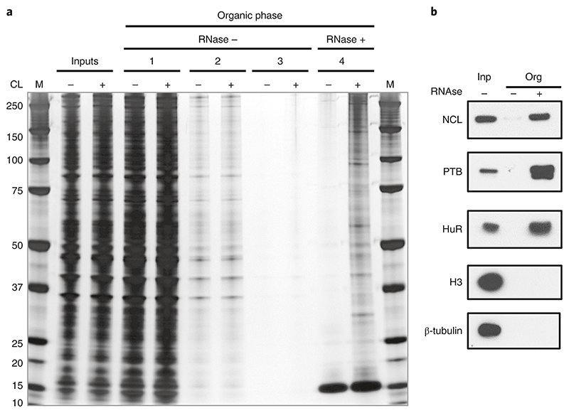
Fig. 6. Silver staining illustrating different stages of the OOPS protocol.
a, The OOPS protocol was performed on non-cross-linked (CL-) and cross-linked (CL+) U2OS cells, and protein samples were obtained from the following stages: cell lysates before separation (inputs), organic phases after each of the first three rounds of TRIzol separation (organic phases 1, 2 and 3) and the organic phases after RNase and final TRIzol separation (organic phase 4). Samples were run, along with a protein ladder (M), on a 4−12% Bis-Tris gel, which was then silver stained. b, Representative western blots for positive RBP controls (NCL; Nucleolin, 100KDa, PTB; 60KDa and HuR; 36KDa) and negative-control proteins (H3; Histone-H3; 18KDa and β-tubulin; 55 KDa). Anti-nucleolin (1:1,000, Clone 4E2, GeneTex, GeneTex, cat. no. GTX13541, RRID:AB_372550); anti-PTB (1:1,000, RRID:AB_2827814); #GTX13541); anti-HuR (1:1,000, Santa Cruz Biotechnology, cat. no. sc-5261, RRID:AB_627770); anti-histone H3 (1:1,000, Bethyl, cat. no. A300−823A, RRID:AB_2118462); and anti-β-tubulin (1:1,000, Cell Signaling Technology, cat. no. 2146, RRID: AB_2210545). Adapted from ref. 5.

