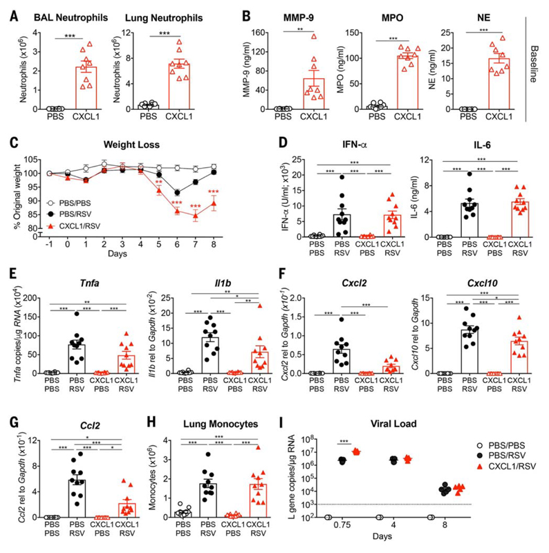
Fig. 6. Mice treated with the neutrophil chemoattractant CXCL1 before RSV infection show an early increase in viral load and develop more severe disease.
Mice were treated with mock (PBS) or 10 μg of CXCL1 intranasally. (A) At 12 hpi, BAL and lung neutrophils were quantified by flow cytometry and (B) MMP-9, MPO, and NE levels in the BAL were quantified by ELISA. Mice were treated with mock (PBS) or 5 to 10 μg of CXCL1 intranasally and after 9 to 12 hours were infected with mock (PBS) or 7.5 × 10⁵ FFUs of RSV intranasally. (C) Weight loss as a percentage of original weight. (D) IFN-α and IL-6 quantified in the BAL using ELISA. Levels of (E) Il1b and Tnfa, (F) Cxcl2 and Cxcl10, and (G) Ccl2 were determined in lung tissue using qPCR. (H) Total number of lung monocytes quantified by flow cytometry as previously described (32). (I) RSV L gene copy numbers were quantified at 0.75, 4, and 8 dpi in lung tissue by qPCR. Data in (A) and (B) are shown as the means ± SEM of eight individual mice per group pooled from two independent experiments. Data in (C) are shown as the means ± SEM of 10 (PBS/PBS) or 14 to 16 (RSV) individual mice pooled from two (PBS/PBS) or three (RSV) independent experiments. Data in (D) to (H) are shown as the means ± SEM of 10 individual mice per group pooled from two independent experiments. Data in (I) are shown as the means ± SEM of five individual mice per group representative of at least two independent experiments, with the dotted line representing the limit of detection of the assay. The statistical significance of differences in (A) and (B) was analyzed using unpaired, two-tailed Student’s t test. The data in (C) and (I) were analyzed using two-way ANOVA with Bonferroni’s post hoc test, and only the statistically significant differences between RSV-infected groups are shown. The data in (D) to (H) were analyzed using one-way ANOVA with Tukey’s post hoc test. *P < 0.05, **P < 0.01, ***P < 0.001.

