Figure 3.
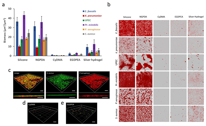
(a) Surface coverage by single species (E. faecalis, K. pneumoniae, E. coli, Pr. mirabilis, P. aeruginosa and S. aureus) biofilms quantified after 72 h incubation on silicone, silver hydrogel, pNGPDA, pCyDMA and p(EGDPEA-co-DEGMA) (labelled EGDPEA) coated silicone catheter segments in AU. Error bars equal ± 1 sd unit, n = 3. (b) The corresponding confocal microscopy images for Syto64 stained E. faecalis, K. pneumoniae, E. coli, Pr. mirabilis, P. aeruginosa and S. aureus growing on each polymer surface. Each image is 160 x 160 μm. (c) 3D representation and transverse view of a dual-species biofilm formed on glass: GFP-tagged S. aureus SH1000 (green) and mCherry labelled P. aeruginosa (red) in a 10:1 ratio. (d)-(e) Three dimensional representation and transverse view showing the lack of mature biofilm on pCyDMA and pEGDPEA. Scale bars represent 50μm.

