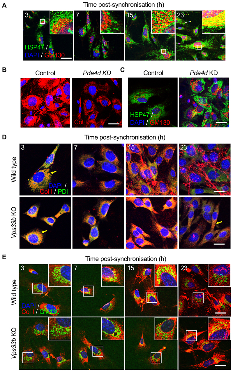
Figure 4. Golgi and post-Golgi transport of PC-I is rhythmic.
(A) Immunofluorescence analysis of collagen chaperone HSP47, and ERGIC and the cis-Golgi (detected by marker GM130) in synchronised MEFs showed gradual co-localisation of HSP47 with GM130 (see insets), n=5. Quantification in Figure S6D. (B) Immunofluorescence analysis shows that PDE4D depletion in MEFs using siRNAs (Pde4d KD) blocked collagen-I secretion into the matrix, which was unaffected in control cells, n=3. (C) Immunofluorescence analysis of HSP47 in Pde4d KD cells show accumulation of HSP47 in GM130 compared to control MEFs, n=5. (D) Immunofluorescence analysis of synchronised iTTFs shows collagen-I secretion (white arrows) was inhibited in CRISPR-mediated Vps33b KO cells, where collagen-I was retained in the ER (PDI staining) (yellow arrows). N=4 biologically independent experiments. (E) Immunofluorescence analysis of synchronised MEFs also shows increased retention of collagen-I occurs in the Golgi (GM130 staining) when Vps33b was knocked out (see insets). N=2 biologically independent experiments. Bars 10 μm.

