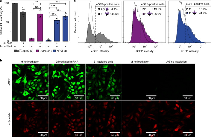
Fig. 5. Light-induced translation in cells.
a, Relative luciferase activity from HeLa cells transfected with differently capped GLuc-mRNAs. The mRNA was capped with the indicated cap analogue. Data of n = 3 independent experiments are shown as mean values ± s.e.m. Statistical significance was determined by two-tailed Student's t-test. Significance levels were defined as *P < 0.05, **P < 0.01, ***P < 0.001. The P value for 1 (+ irr. cells, − irr. mRNA) versus 1 (− irr. cells, − irr. mRNA) is 1.26 × 10⁻³. The P value for 2 (+ irr. cells, − irr. mRNA) versus 2 (− irr. cells, − irr. mRNA) is 3.53 × 10⁻⁴. The P value for 2 (− irr. cells, + irr. mRNA) versus 2 (− irr. cells, − irr. mRNA) is 1.52 × 10⁻⁴. b, Confocal laser scanning microscopy images of HeLa cells co-transfected with differently capped eGFP-mRNAs and cap 0-mScarlet-I-mRNA. mRNAs contain m5C and m1Ψ. AG: ApppG-capped mRNA represents cap-independent translation. (0): m7GpppG-capped eGFP-mRNA. 2: NPM capped-eGFP-mRNA, either non-irradiated, irradiated in cells or irradiated before transfection (irradiated mRNA). The top row shows the eGFP fluorescence and the bottom row the mScarlet-I fluorescence. Scale bars, 50 µm. For all images, background subtraction was performed with ImageJ (30 pixels). Shown is one representative set of n = 3 independent experiments. c, Flow cytometry of HeLa cells transfected with differently capped mRNAs (with cap analogues 0, 1, 2 or ApppG (AG)). Untransfected cells are set as gate for eGFP-negative cells. Irradiation is indicated by an LED icon. Shown is one representative measurement of n = 3 independent experiments.

