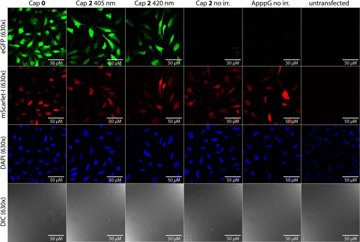
Extended Data Fig. 3. Representative 630x magnification confocal microscopy images of irradiated (405 nm, 420 nm) and non-irradiated HeLa cells transfected with eGFP- and mScarlet-I-mRNA with DAPI staining.
HeLa cells were transfected with differently capped eGFP-mRNA containing m5C and m1Ψ and m7GpppG-capped mScarlet-I-mRNA containing m5C and m1Ψ. Untransfected cells served as control. ApppG-capped mRNA represents cap-independent translation. The m7GpppG-capped eGFP-mRNA (0) served as positive control. The NPM-(2) caged eGFP-mRNA was either not irradiated or irradiated in cells (405 nm, 60 s/420 nm 180 s). The top two rows show the 630x magnification (63x objective) of the red channel (mScarlet-I) or the green channel (eGFP) while the bottom two show the DAPI staining and DIC channel. Shown is one representative experiment of three independent experiments (n = 3).

