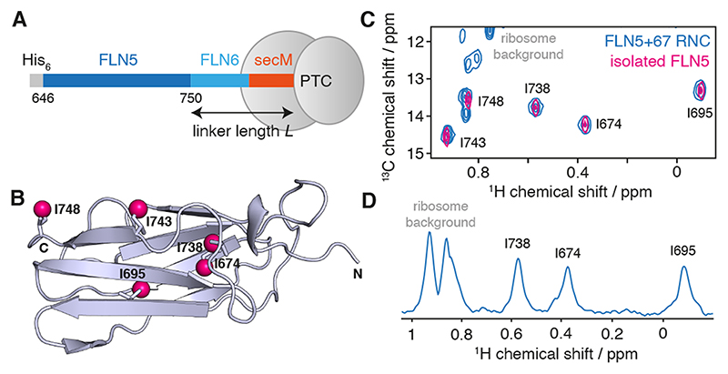
Figure 1. Solution-state NMR spectroscopy of FLN5+6 RNCs.
(A) Design of RNC constructs [9], showing the complete FLN5 filamin domain (646–750) flanked by an N-terminal His6 affinity tag, and the subsequent FLN6 domain and 17 amino acid secM arrest motif, of total length L. (B) Crystal structure (1qfh [15]) of FLN5 showing the location of the five isoleucine residues, with Cδ atoms highlighted as magenta spheres. (C) 1H,13C HMQC correlation spectrum of a [2H,13CH3-Ile]-labelled FLN5+67 RNC (blue) and isolated [2H,13CH3-Ile]-labelled FLN5 (298 K, 950 MHz). (D) 1D spectrum showing the first increment of an 1H,13C HMQC measurement of the FLN5+67 RNC.

