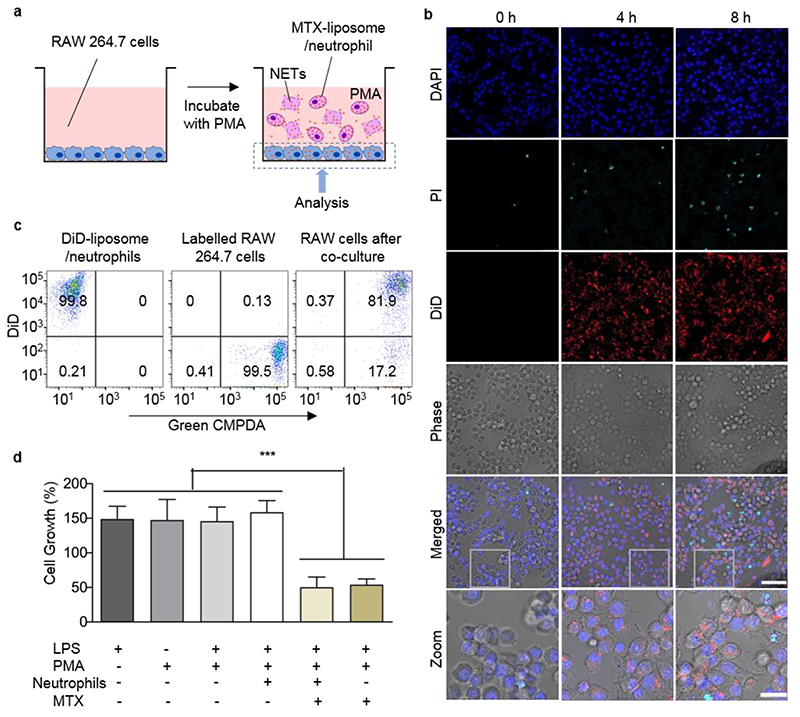
Figure 3. Neutrophil-mediated delivery and effect of MTX-liposome on co-cultured macrophages (RAW 264.7 cells).
a, Schematic illustration of the in vitro co-culture system of macrophages (RAW 264.7 cells) and MTX-liposome/neutrophils. b, CLSM images of RAW 264.7 cells after incubation with liposome/neutrophils at 0 h, 4 h and 8 h in presence of PMA (controls without PMA are in SI, Figure S11). The nuclei of RAW 264.7 cells were stained with DAPI, the released DNA fragments of neutrophils were stained with PI and the liposomes were labelled with DiD. Scale bar: 50 μm. Zoom images, Scale bar: 10 μm. c, Flow cytometry analysis of RAW 264.7 cells after co-culture with DiD-liposome/neutrophils. Green CMPDA channel shows RAW 264.7 cell labelling and the DiD channel represents liposome labelling. d, Proliferation of RAW 264.7 cells after treatment with inflammatory cytokines and co-culture with MTX-liposome/neutrophils compared to basal medium. Cell viability was measured using Cell Counting Kit-8 (mean ± s.d., n= 3 independent experiments). ***P < 0.001, one-way ANOVA, Bonferroni post hoc test.

