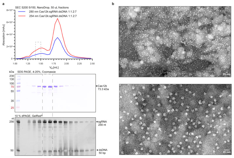
Extended Data Fig. 1. ShCas12k-sgRNA-target DNA complex assembly.
a, Top: Size exclusion chromatography analysis of the ShCas12k-sgRNA-target DNA complex. Middle: SDS-PAGE analysis of fractions from a. Proteins were visualised by Coomassie blue staining. Bottom: denaturing PAGE analysis of fractions from a. Nucleic acids were stained with a fluorescent dye (Gel Red). b, Representative negative stain EM micrographs of the ShCas12k-sgRNA-dsDNA complex at 68,000x magnification (top) and 180,000x magnification (bottom). Experiment was repeated three times independently with similar results. For gel source data, see Supplementary Figure 1.

