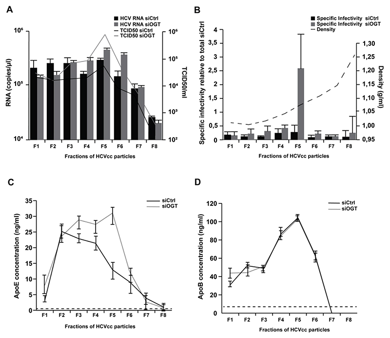
Figure 5. Silencing of OGT modulates HCVcc biophysical properties.
(A) Separation of HCVcc by iodixanol density gradient ultracentrifugation. HCVcc were produced in non-targeting siRNA control- or siOGT-transfected Huh7.5.1 cells. After overlaying HCVcc (JcR2A) on a 4%-40% iodixanol step gradient and ultracentrifugation for 16h, fractions of HCV particles were used to infect naïve Huh7.5.1 cells in order to determine TCID50. HCV RNA of each fraction was purified and analyzed by RT-qPCR. Data are expressed as mean ± s.d. from three independent experiments. (B) Specific infectivity (TCID50/RNA) was calculated and the density was determined by weighting each fraction. Specific infectivity of each fraction is expressed as fold change as compared to the total infectivity of the control. Data are expressed as mean ± s.d. from three independent experiments. (C-D) ApoE and ApoB concentrations in the individual fractions were determined by ELISA. The dashed lines indicate limits of quantification of the assays. Data are expressed as mean ± s.d. from three independent experiments.

