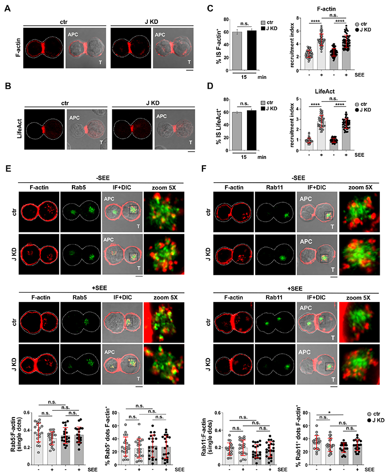
Figure 5. BBS1 is dispensable for synaptic F-actin accumulation and endosomal F-actin dynamics.
(A,B) Immunofluorescence analysis of F-actin in control and BBS1KD (J KD) Jurkat cells (A), or control and BBS1KD (J KD) Jurkat cells transiently transfected with the LifeAct reporter (B), and conjugated with SEE-loaded or with unloaded Raji cells (APCs) for 15 min. Representative images (medial optical sections) of the conjugates formed in the presence of SEE are shown (see Fig.4G,H for SEE-independent conjugates). (C,D) Left, Quantification (%) of 15-min SEE-specific conjugates harboring F-actin (C) and LifeAct (D) staining at the IS (≥25 cells/sample, n≥3, unpaired two-tailed Student’s t-test). Right, Relative F-actin (C) or LifeAct (D) fluorescence intensity at the IS in 15-min conjugates of control (ctr) and BBS1KD (J KD) Jurkat cells with Raji cells in the absence or presence of SEE (≥15 cells/sample, n=3, Kruskal-Wallis test). (E,F) Immunofluorescence analysis of endosomal F-actin in 15-min conjugates of control (ctr) and BBS1KD (J KD) Jurkat cells with Raji cells (APCs) in the absence or presence of SEE. Conjugates were costained for Rab5 (E) and Rab11 (F) (see Fig.S4C for mask generation). Bottom, Colocalization of F-actin on individual dots (left) and quantification of Rab5+/Rab11+ dots positive for F-actin+(right) (10 cells/sample, 15 dots/cell, n=2, Kruskal Wallis test or One-way ANOVA based on normality distribution of different data sets). Size bar, 5 μm. The data are expressed as mean±SD. ****P≤0.0001; n.s., not significant.

