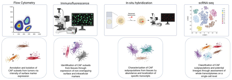
Figure 2. Identification of discrete CAF subsets achieved through different experimental systems.
From left to right: Flow cytometry has been employed to annotate and sort different CAF subgroups from tumors, also enabling CAF subset isolation for further experimentation into CAF phenotypes and tasks. Immunofluorescence and RNA in-situ hybridization experiments revealed CAF subsets from tissue samples, based on discrete expression of surface and intracellular protein markers and transcripts; these methods also reveal important spatial information regarding CAF subtype localization in the TME. Finally, scRNA sequencing has provided a breakthrough in stratification of a multitude of novel CAF subtypes through high resolution characterization of whole transcriptomes on a single cell level, providing information on rare CAF subsets and potential information regarding CAF lineages. The figure was created with BioRender.com.

