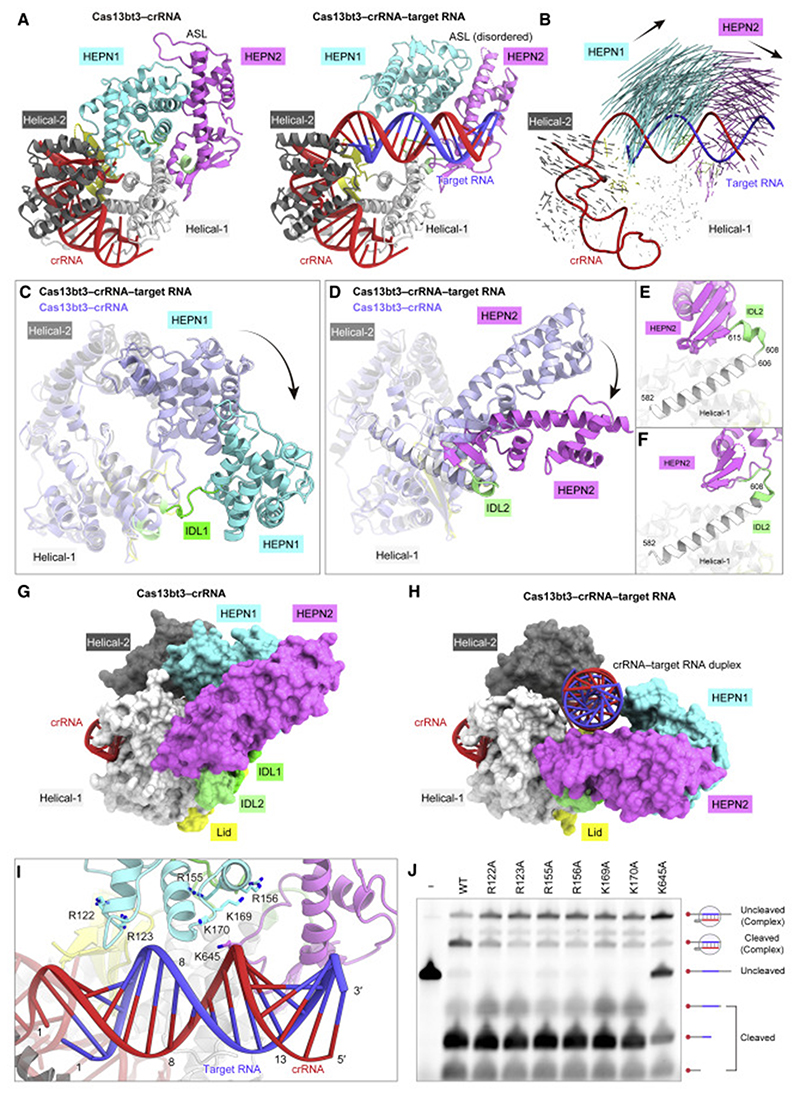
Figure 5. Structural differences between the Cas13bt3 binary and ternary complexes.
(A) Structural comparison of the Cas13bt3 binary (left) and ternary (right) complexes.
(B) Structural transition between the binary and ternary complexes. Vectors indicate the transitions of equivalent Cα atoms between the binary and ternary complexes.
(C and D) Structural changes in the HEPN1 (C) and HEPN2 (D) domains between the binary and ternary complexes.
(E and F) Conformational transitions of IDL2 between the binary (E) and ternary (F) complexes.
(G and H) Surface representations of the binary (G) and ternary (H) complexes.
(I) Basic loop regions in the vicinity of the crRNA-target RNA duplex.
(J) In vitro RNAc leavage activities of the wild-type and mutant Cas13bt3s. The 50-Cy5-labeled target RNA, containing the 30-nt target sequence, was incubated with the Cas13bt3-crRNA complex for 60 min and then analyzed by denaturing urea PAGE.

