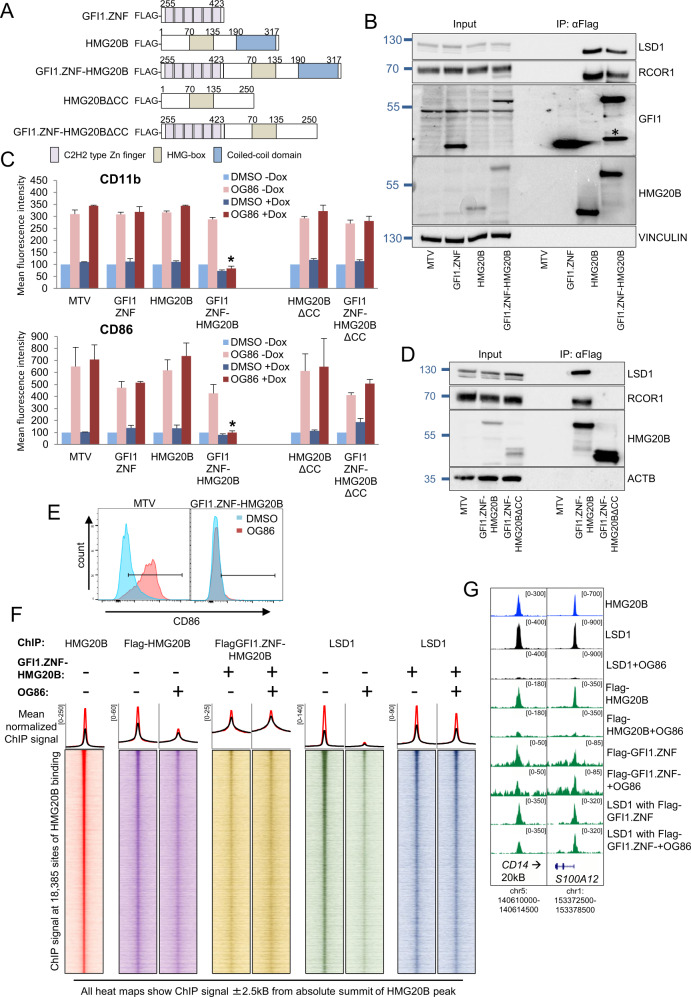
Fig. 4. Functional recruitment of LSD1 to chromatin by a GFI1 DNA-binding domain:HMG20B fusion protein.
A Construct maps. B Western shows doxycycline-induced expression of the indicated constructs and αFlag IPs in THP1 AML cells. * Indicates degradation product. C THP1 AML cells were infected with lentiviruses expressing the indicated doxycycline-inducible (Dox) GFI1 fusion or control constructs and then treated with 250 nM OG86 or DMSO vehicle. Bar graphs indicate means + SEM fluorescence intensity for the indicated markers, as determined by flow cytometry, in the indicated conditions 24 h later (n = 3). * indicates P < 0.05 for the indicated condition versus all other OG86 + Dox conditions by one-way ANOVA and a Tukey post hoc test. D Western blot shows Dox-induced expression of the indicated constructs and αFlag IPs in THP1 AML cells. E Representative flow cytometry plots. F Heatmaps show ChIP signal for the indicated proteins and conditions. Line graphs above show mean normalized ChIP signal for each heatmap surrounding all (black line) or the strongest 20% (red line) of HMG20B peaks. G Exemplar ChIPseq peaks tracks.

