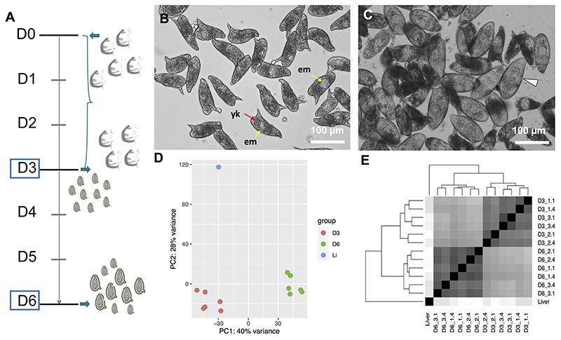
Figure 1.
(A) Timeline depicting the experimental design. On day 0 (D0) adult worms perfused from infected mice were washed and placed in culture for three days. The worms laid eggs, i.e. in vitro laid eggs (IVLE), for three days (bracket). On day 3 (D3) the worms were removed from the culture, and half of the IVLE were collected for RNAseq - D3, early embryogenesis sample. The remaining IVLE were cultured for three more days, and on day 6 (D6) they were collected for RNAseq - D6, late embryogenesis sample. (B, C) Representative micrographs of 3 days old- (B) and 6 days old- (C) in vitro laid eggs (IVLE). Scale bar: 100 μm. em, embryo (yellow arrow); yk, yolk (red arrow); white arrowhead, fully developed egg containing the mature miracidium. (D) Clustering of egg samples using Principal Component Analysis. D3, D3 IVLE; D6, D6 IVLE; Li, liver eggs (E) Clustering of egg samples using Sample Distance Matrix. Names of samples are described in Supplementary Table S1.

