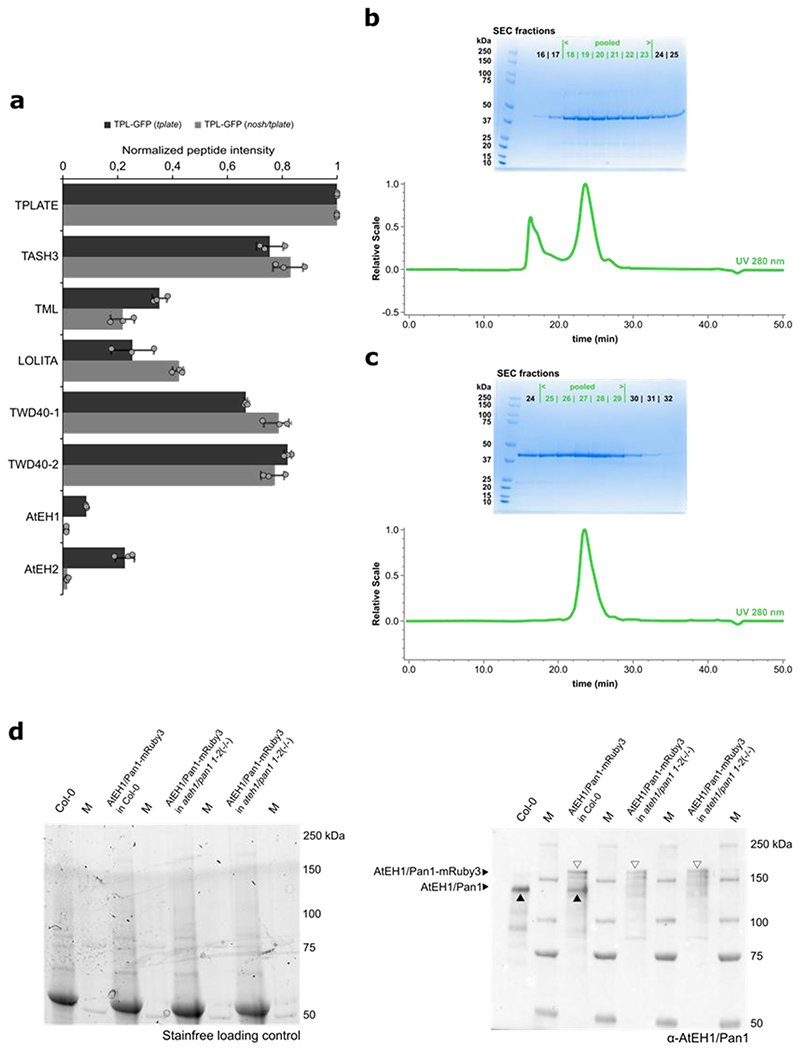
Extended Data Fig. 3. Comparative interactomics and AtEH1/Pan1 antibody specification.
a) Graph depicting the normalized peptide intensities of TPC subunits obtained from MS analysis. For each TPC subunit, the intensities of only those peptides that were present in all experiments (for both baits and in all replicas) were averaged and normalized to the values of the corresponding bait protein. Error bars correspond to ± SD and are based on three technical repeats. The results show that nosh does not affect the hexameric TPC formation, but that the association with the AtEH/Pan1 proteins is weakened. b) Coomassie stained gel of the obtained SEC fractions and a quality control HPLC analysis performed using a Superdex 200 increase 10/300 for the batch of recombinant AtEH1 C-term fragment used for rabbit immunization. Fractions 18 - 23 were pooled to immunize (marked in green). c) Coomassie stained gel of the obtained SEC fractions and a quality control HPLC analysis performed using a Superdex 200 increase 10/300 for the batch used for antibody purification. Fractions 25 - 29 were pooled for purification (marked in green). d) Stain free gel and blot for testing AtEH1/Pan1 antibody specificity. In the Col-0 sample only the native AtEH1/Pan1 band is prominently observed (marked with a black arrowhead), while in the pH3.3::AtEH1/Pan1-mRuby3 (Col-0) sample, both the native and the transgene fusion protein (marked with a white arrowhead) can be observed. Two homozygous pH3.3::AtEH1/Pan1-mRuby3 (ateh1/pan1 1-2 -/-) lines show only the transgene fusion protein (marked with white arrowhead).

