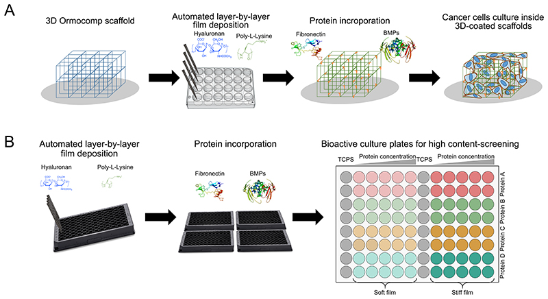
Figure 1. Schematic representation of the overall process of microtumor development on 3D bioactive scaffolds.
(A) Polyelectrolyte multilayer film coating using a liquid handling robot, protein loading and cell seeding on the 3D Ormocomp scaffold and (B) Polyelectrolyte multilayer coating and protein loading on the 96-well plates for high-throughput cellular assays.

