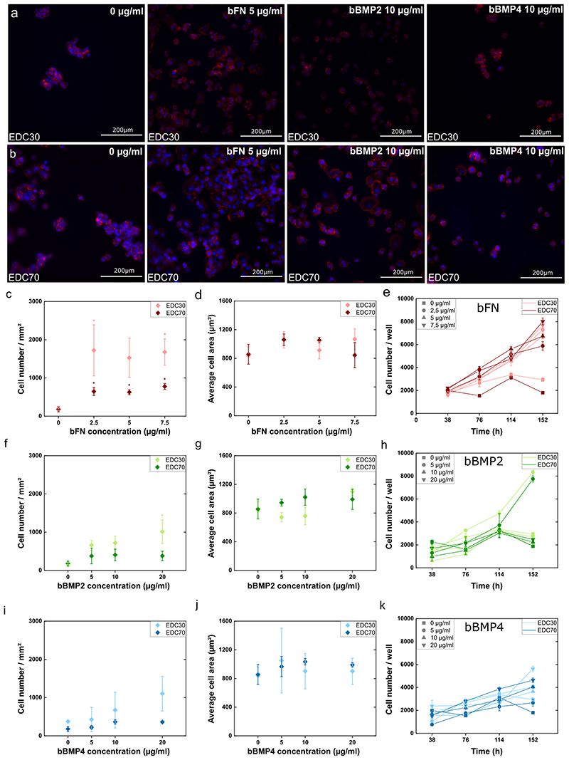
Figure 4. Cell adhesion, spreading and proliferation of PANC1 on soft (EDC30) and stiff (EDC70) PEM film.
Fluorescence images of cells with nucleus labelled with DAPI (blue) and actin cytoskeleton labelled with rhodamine (red) on (A) soft film and (B) stiff film with FN, bBMP2 or bBMP4. Cell adhesion (C, F, I), cell spreading (D, G, J), and proliferation (E, H, K) were quantified in the presence of fibronectin (C-E), BMP2 (F-H), or BMP4 (I-K). The mean of 3 independent experiments is plotted with SEM. * indicates that the value is significantly different from the control condition.

