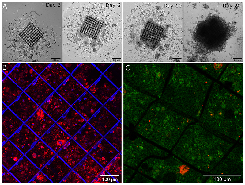
Figure 7. 3D culture of PANC1 cells on the bioactive scaffold loaded with 5 μg/ml fibronectin.
(A) Bright field images of cells after 3, 4, 10 and 20 days of culture. Confocal images of PANC1 cells (B) with nucleus stained blue (DAPI) and cytoskeleton stained red (rhodamine) after 11 days in culture and (C) Live & Dead assay after 20 days in culture with live cells labelled in green and dead cells labelled in red.

