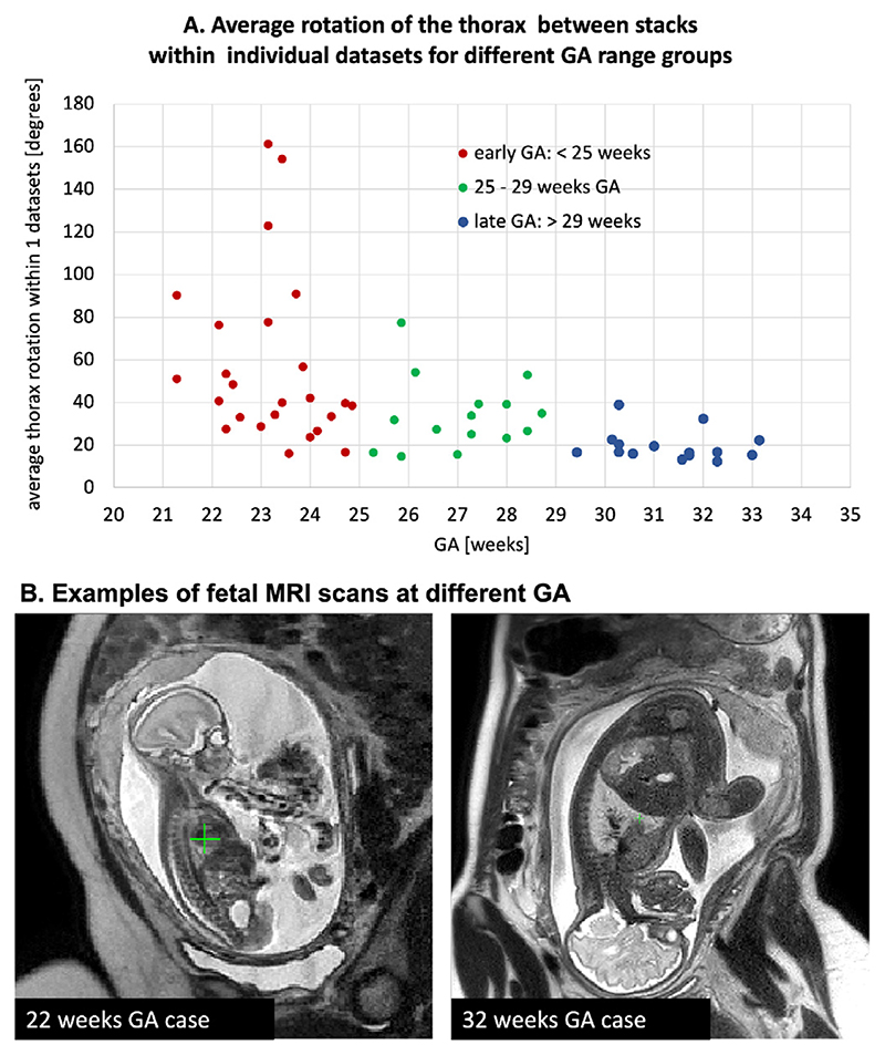
Fig. 3.
A. Comparison of the degree of the global fetal mobility during MRI acquisition for 55 randomly selected datasets acquired at St. Thomas’s Hospital and Evelina London Children’s Hospital using the same acquisition protocol: average rotation ranges for the fetal thorax ROI between stacks within individual datasets. It includes < 25 weeks GA early (red), 25–29 weeks GA (green), > 29 weeks GA late (blue) groups. B. Examples of fetal MRI scans at 22 and 32 weeks GA.

