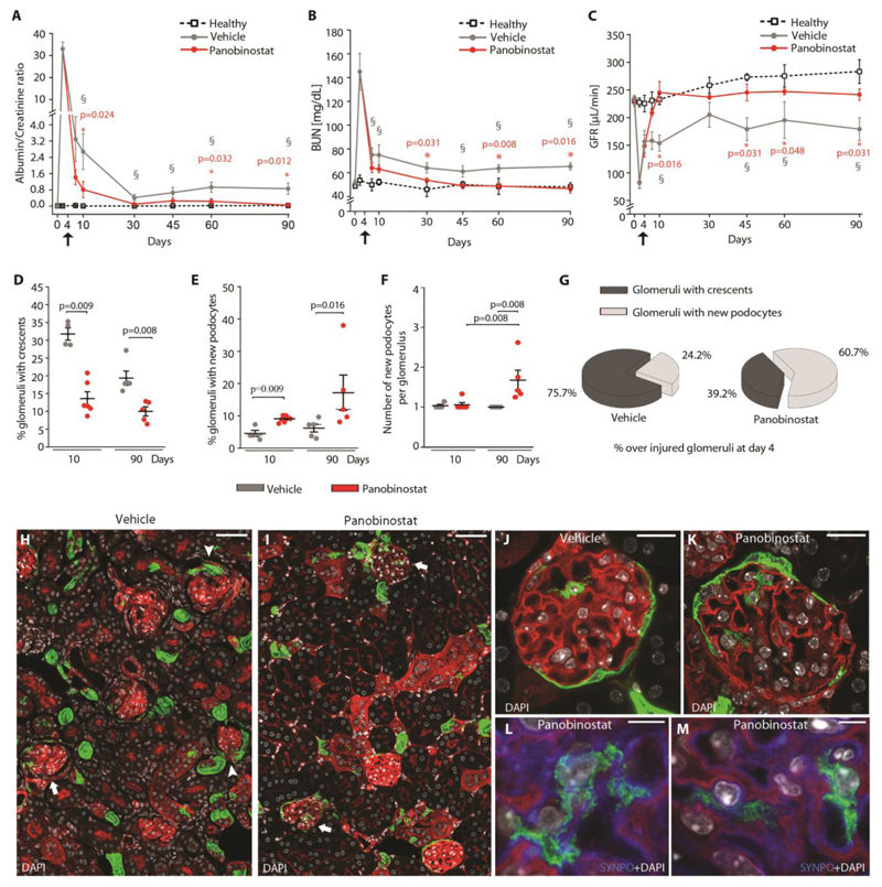
Fig. 4. Panobinostat ameliorates crescentic glomerulonephritis and prevents kidney failure.
(A) Time course of urine albumin/creatinine ratio determined in healthy (n=4), vehicle (n=9 at day 4 and 10, n=5 at day 30, 45, 60 and 90) and panobinostat (n=11 at day 4 and 10, n=5 at day 30, 45, 60 and 90) treated mice. (§) Significance of vehicle versus healthy mice: day 10 p=0.004, day 30, 45, 60 and 90 p=0.016). (*) Significance of panobinostat versus vehicle. (B) Time course of BUN determined in healthy (n=4), vehicle (n=9 at day 4 and 10, n=5 at day 30, 45, 60 and 90) and panobinostat (n=11 at day 4 and 10, n=5 at day 30, 45, 60 and 90) treated mice. (§) Significance of vehicle versus healthy mice: day 7, 10 p=0.019, day 45, 60 and 90 p=0.032). (*) Significance of panobinostat versus vehicle. (C) GFR determined in healthy (n=4), vehicle (n=5) and panobinostat (n=4) treated mice. Black arrows indicate the starting point for drug treatment. Data are mean ±SEM. (§) Significance of vehicle versus healthy mice: day 10, 45 and 90 p=0.016, day 60 p=0.032). (*) Significance of panobinostat versus vehicle. (D) Percentage of glomeruli presenting crescents at day 10 (vehicle n=4, panobinostat n=6) and day 90 (vehicle n=5, panobinostat n=5). (E) Percentage of glomeruli with new podocyte(s) over total number of glomeruli at day 10 (vehicle n=4, panobinostat n=6) and day 90 (vehicle n=5, panobinostat n=5). (F) Dot plot showing the mean number of Pax2+ cells observed within glomeruli at day 10 (vehicle n=4, panobinostat n=6) and day 90 (vehicle n=5, panobinostat n=5). (G) Percentage of glomeruli with crescent and glomeruli with new podocytes observed at day 90 (vehicle n=5, panobinostat n=5). Percentages were calculated among glomeruli presenting crescent at day 4 after injury. (H, I) Representative images of kidneys from vehicle (H) and panobinostat (I) treated Pax2.rtTA;TetO.Cre;R26.mT/mG mice sacrificed at day 90, showing the presence of Pax2+ derived cells inside the glomerular tuft in panobinostat treated mice (arrows) and the presence of crescent in vehicle treated mice (arrowheads). Bars=50 μm. (J, K) Representative image of a glomerulus showing Pax2+ derived cells inside the glomerular tuft in vehicle (J) and panobinostat (K) treated Pax2.rtTA;TetO.Cre;R26.mT/mG mice sacrificed at day 90. Bars=20 μm. (L, M) Higher magnification showing foot processes and synaptopodin (SYNPO) expression (blue) in the Pax2+ derived cell in panobinostat treated Pax2.rtTA;TetO.Cre;R26.mT/mG mice sacrificed at day 90. DAPI counterstains nuclei (white). Bars=5 μm.
Signals for fluorescent mT/mG proteins are GFP, green, and tdTomato, red. In dot plots (D-F) bars indicate mean values. Individual scores are shown. Statistical significance in A-F was calculated by Mann-Whitney test. Numbers on graph represent p-values.
BUN, blood urea nitrogen; DAPI, 4',6-diamidino-2-phenylindole; SYNPO, synaptopodin.

