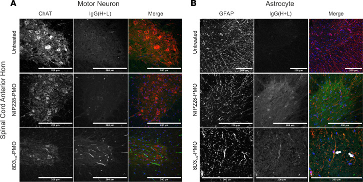
Figure 6. Cellular localization of 8D3130-PMO to the astrocytes in the spinal cord.
Representative confocal images of spinal cord following single 50 mg/kg administration of 8D3130-PMO and NIP228-PMO. The spinal cord was isolated from adult mice 24 hours postadministration following perfusion fixation. (A) Motor neurons (ChAT) in the anterior horn of the spinal cord, and 8D3130-PMO identified by human secondary antibody, IgG(H+L), showed no overlap (merge). Fluorescence indicated a retention of the 8D3130-PMO [IgG(H+L)] in the vasculature. (B) Astrocytes of the anterior gray horn (GFAP) were colocalized with 8D3130-PMO [IgG(H+L)] (arrowheads). Scale bar represents 200 μm.

